Author: Admin

  • TermDefinition
    Biological ClassificationThe systematic arrangement of living organisms into groups based on similarities and evolutionary relationships.
    TaxonomyThe science of naming, describing, and classifying organisms.
    SystematicsThe study of biological diversity and the evolutionary relationships among organisms.
    Binomial NomenclatureA universal naming system assigning each species a two-part name: genus and species.
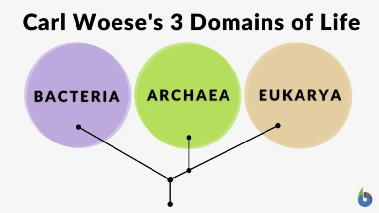
    Taxonomic HierarchyThe ordered ranking of taxa from broad to specific: domain, kingdom, phylum, class, order, family, genus, species.

    Biological classification provides a framework for organising the immense diversity of life on Earth. By grouping organisms based on shared features and genetic relationships, scientists can communicate effectively, make predictions about unknown species, and understand evolutionary history. The system originated with Carl Linnaeus’s morphological approach and has since evolved to incorporate molecular data, making classification more accurate and reflective of true phylogenetic relationships.

    • Organises biodiversity into manageable and meaningful groups.
    • Enables clear communication among scientists worldwide through standardised naming.
    • Provides insights into evolutionary relationships between organisms.
    • Helps predict characteristics of newly discovered organisms.
    • Supports conservation efforts by identifying species and their roles in ecosystems.
    • Facilitates research by grouping organisms with similar traits.

    🧠 Examiner Tip: Always italicise genus and species names and capitalise only the genus when writing binomial nomenclature (e.g., Homo sapiens).

    • Based on observable characteristics and genetic similarities.
    • Uses a hierarchical system of ranks from broad to specific.
    • Includes both morphological and molecular evidence.
    • Reflects evolutionary history wherever possible.
    • Employs binomial nomenclature for universal identification.
    • Allows for flexibility and revision as new data emerge.

    🧬 IA Tips & Guidance: A possible IA could compare species identification using morphological keys versus DNA barcoding techniques.

    • Artificial systems: Based on one or few characteristics, often for convenience (e.g., habitat or size).
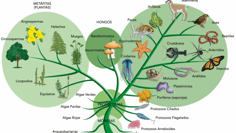
    • Natural systems: Based on multiple characteristics reflecting evolutionary relationships.
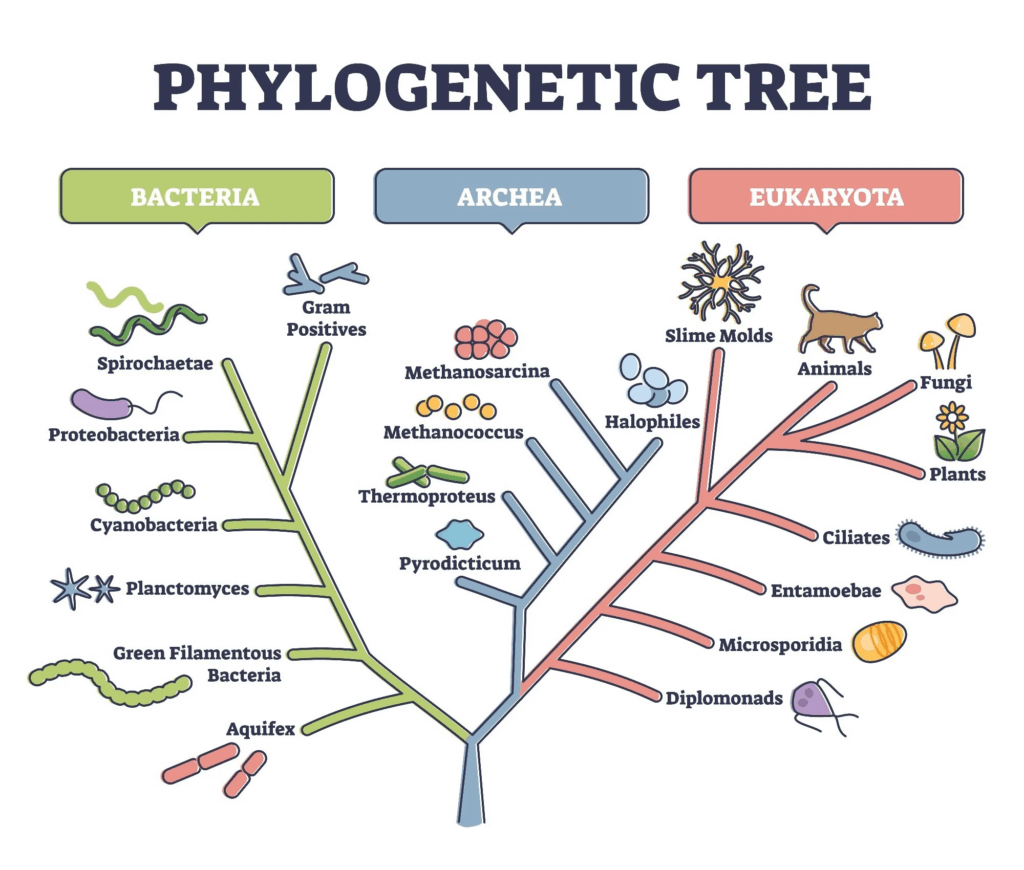
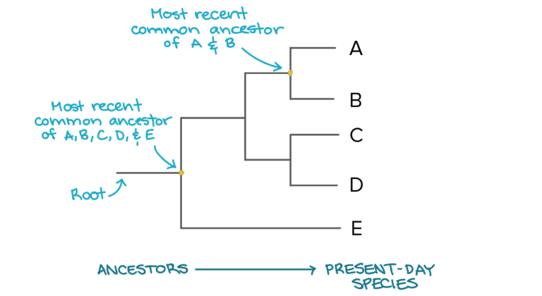
    • Phylogenetic systems: Focused on common ancestry using morphological and molecular data.
    • Systems can shift as new technologies and evidence become available.
    • Phylogenetic classification is currently the most accepted method.
    • Fossil evidence can be integrated into classification.

    🌐 EE Focus: An EE could analyse how molecular evidence has reshaped classification within a specific group of organisms.

    • Dichotomous keys for identification.
    • Field guides for local species.
    • Microscopy for cellular characteristics.
    • DNA sequencing for molecular comparisons.
    • Bioinformatics for analysing large datasets.
    • Phylogenetic software for tree construction.

    ❀️ CAS Link: A CAS project could involve creating a field guide for local biodiversity, integrating both scientific names and common names.

    • Morphological similarities may be due to convergent evolution, not shared ancestry.
    • Genetic data may conflict with traditional classification.
    • Hybridisation can blur species boundaries.
    • Cryptic species may remain undetected without molecular analysis.
    • Classification is dynamic β€” constant updates are required.
    • Human bias can influence early classification decisions.

    πŸ” TOK Perspective: Classification systems reflect human attempts to impose order on nature; changes in technology and perspective can reshape these systems over time.

    🌍 Real-World Connection:
    Accurate classification underpins agriculture, medicine, conservation, and environmental policy by ensuring correct species identification.

    πŸ“ Paper 2:
    Be ready to define taxonomy and systematics, outline the hierarchy of classification, and compare artificial, natural, and phylogenetic systems.

  • TermDefinition
    BiodiversityThe variety of life at all levels, from genes to ecosystems.
    PhylogeneticsThe study of evolutionary relationships among organisms.
    CladogramDiagram showing evolutionary relationships based on shared derived traits.
    Molecular ClockA method estimating evolutionary time based on mutation rates in DNA or protein sequences.
    Convergent EvolutionThe independent evolution of similar features in unrelated lineages due to similar environmental pressures.

    Evolutionary relationships underpin modern biology, explaining how species are connected through common ancestry. Biodiversity is shaped by evolutionary processes, environmental changes, and ecological interactions. Studying phylogenetics allows scientists to reconstruct the tree of life, identifying shared ancestry and divergence events. Advances in DNA sequencing and bioinformatics have transformed our understanding of biodiversity, revealing cryptic species and clarifying relationships that morphological studies alone could not resolve. Conservation biology increasingly depends on evolutionary data to prioritise species and habitats for protection.

    • Morphological analysis: comparing structures to infer relationships.
    • Molecular phylogenetics: uses DNA, RNA, or protein sequences to build evolutionary trees.
    • Molecular clocks: estimate divergence times based on mutation rates.
    • Cladistics: groups organisms by shared derived traits (synapomorphies).
    • Fossil calibration: uses fossil ages to anchor phylogenetic timelines.
    • Bioinformatics: computational tools for large-scale sequence analysis.

    🧠 Examiner Tip: When interpreting cladograms, remember that branch length may or may not indicate evolutionary time β€” check the diagram’s scale.

    • Divergent evolution: species from a common ancestor become more different over time.
    • Convergent evolution: unrelated species evolve similar traits due to similar selection pressures.
    • Parallel evolution: related species evolve similar traits independently.
    • Adaptive radiation: rapid diversification from a common ancestor into multiple ecological niches.
    • Coevolution: two or more species reciprocally influence each other’s evolution.
    • Extinction events: can reset evolutionary trajectories and open niches.

    🧬 IA Tips & Guidance: A bioinformatics-based IA could compare mitochondrial DNA sequences to infer evolutionary relationships between local species.

    • Genetic diversity: variation in genes within a population.
    • Species diversity: number and relative abundance of species in an area.
    • Ecosystem diversity: variety of habitats and ecological processes.
    • Biodiversity supports ecosystem stability and resilience.
    • High biodiversity often correlates with ecosystem productivity.
    • Conservation strategies target all three diversity levels.

    🌐 EE Focus: An EE could investigate how genetic diversity influences species survival in changing environments, using molecular data for analysis.

    • Habitat loss from agriculture, urbanisation, and deforestation.
    • Overexploitation of species for food, medicine, or trade.
    • Pollution affecting ecosystems and species health.
    • Climate change altering habitats and migration patterns.
    • Invasive species outcompeting native organisms.
    • Conservation biology aims to mitigate these impacts using evolutionary insights.

    ❀️ CAS Link: A CAS project could involve participating in biodiversity surveys and using phylogenetic tools to classify observed species.

    • Phylogenetic diversity can be used to prioritise species conservation.
    • Protecting evolutionarily distinct species preserves unique traits.
    • Genetic data reveal population structure for targeted management.
    • Evolutionary history informs restoration ecology projects.
    • Endangered species lists increasingly consider evolutionary uniqueness.
    • Conservation decisions may integrate both ecological and evolutionary data.

    πŸ” TOK Perspective: The use of molecular data in biodiversity studies highlights how changing tools and technologies shape our understanding of the natural world and influence policy.

    🌍 Real-World Connection:
    Understanding evolutionary relationships is vital for predicting disease spread, managing endangered species, and sustaining ecosystems under climate change.

    πŸ“ Paper 2:
    Be ready to construct or interpret phylogenetic trees, compare patterns of evolution, and explain how evolutionary knowledge supports biodiversity conservation.

  • TermDefinition
    DomainThe highest taxonomic rank, grouping all life into broad categories based on fundamental genetic and cellular differences.
    ArchaeaDomain of prokaryotic organisms distinct from bacteria, often living in extreme environments.
    BacteriaDomain of prokaryotic organisms with diverse metabolisms, including many important in ecology and medicine.
    EukaryaDomain containing all organisms with eukaryotic cells (nucleus and membrane-bound organelles).
    KingdomTaxonomic level below domain, grouping organisms with shared fundamental characteristics.

    The classification of life into domains and kingdoms reflects the deepest evolutionary divisions among organisms. The three-domain system β€” Bacteria, Archaea, and Eukarya β€” is based on molecular and genetic evidence, particularly ribosomal RNA sequences. Within each domain, organisms are further divided into kingdoms, reflecting key structural, metabolic, and evolutionary traits. This system captures both ancient divergences in life’s history and the diversity of forms and functions that have evolved since.

    • Bacteria: Prokaryotes with peptidoglycan cell walls, circular DNA, and diverse metabolisms. Examples: Escherichia coli, Streptococcus.
    • Archaea: Prokaryotes lacking peptidoglycan, often extremophiles (thermophiles, halophiles, methanogens). Unique membrane lipids and gene expression mechanisms.
    • Eukarya: All organisms with eukaryotic cells, including protists, fungi, plants, and animals.
    • Domain differences are supported by rRNA sequence comparisons and molecular biology evidence.
    • Domains represent the most ancient evolutionary splits, over 3 billion years ago.
    • Archaea and Eukarya share more recent common ancestry than either does with Bacteria.

    🧠 Examiner Tip: In domain comparisons, always mention cell wall composition, ribosome type, and rRNA sequence differences.

    • Domain Bacteria: Often grouped into one kingdom (Eubacteria).
    • Domain Archaea: Often grouped into one kingdom (Archaebacteria).
    • Domain Eukarya: Contains multiple kingdoms:
      • Protista: Mostly unicellular eukaryotes, diverse in form and nutrition.
      • Fungi: Multicellular or unicellular heterotrophs with chitin cell walls.
      • Plantae: Multicellular autotrophs with cellulose cell walls, photosynthetic.
      • Animalia: Multicellular heterotrophs without cell walls, complex body plans.
    • Modern research suggests Protista is paraphyletic, with groups reclassified based on molecular data.
    • Kingdom classification can vary depending on system used (e.g., 5-, 6-, or more kingdoms).

    🧬 IA Tips & Guidance: A possible IA could investigate environmental conditions affecting growth rates of representative organisms from different domains.

    • Bacteria: Binary fission reproduction, 70S ribosomes, sensitive to traditional antibiotics.
    • Archaea: Resistant to most antibiotics, unique lipid monolayers in membranes, ability to survive extreme conditions.
    • Eukarya: Mitotic and meiotic cell division, 80S ribosomes in cytoplasm, membrane-bound organelles.
    • Fungi: Absorptive nutrition, decomposers, important in symbiosis (mycorrhizae).
    • Plantae: Photosynthetic autotrophs, alternation of generations life cycle.
    • Animalia: Nervous and muscular systems for rapid response and movement.

    🌐 EE Focus: An EE could compare molecular phylogenies of organisms from different domains to test evolutionary relationships suggested by rRNA evidence.

    • Carl Woese introduced the three-domain system in the late 20th century.
    • Based on sequencing of the 16S and 18S rRNA genes.
    • Revealed Archaea are genetically closer to Eukarya than to Bacteria.
    • Supports the idea of a universal common ancestor.
    • Highlights the importance of molecular data in reshaping classification.
    • Suggests early life experienced horizontal gene transfer, complicating lineage tracing.

    ❀️ CAS Link: A CAS activity could involve creating an illustrated timeline of the evolutionary emergence of domains and kingdoms for a school biology exhibition.

    • Medical: distinguishing between bacterial and archaeal pathogens for correct treatment.
    • Environmental: identifying microbial communities in extreme environments.
    • Evolutionary research: tracing the origin of complex cellular features.
    • Conservation: cataloguing biodiversity for protection.
    • Biotechnology: selecting species with useful enzymes (e.g., thermostable DNA polymerase from Thermus aquaticus in Archaea).
    • Education: simplifying biological diversity into a teachable structure.

    πŸ” TOK Perspective: The division of life into domains shows how advances in technology (DNA sequencing) can reshape scientific classification systems and challenge traditional frameworks.

    🌍 Real-World Connection:
    Domain and kingdom classification helps scientists track emerging diseases, identify extremophiles for biotechnology, and guide conservation of biodiversity.

    πŸ“ Paper 2:
    Be ready to compare domains in a table, give examples from each kingdom, and explain molecular evidence supporting the three-domain system.

  • TermDefinition
    TaxonomyThe science of classifying organisms into groups based on shared characteristics.
    Binomial NomenclatureThe two-part scientific naming system for species, consisting of genus and species names.
    Taxon (plural: taxa)A group of organisms classified together at any level in the taxonomic hierarchy.
    PhylogenyThe evolutionary history and relationships among species.
    CladisticsA classification method that groups organisms by common ancestry using shared derived characteristics.

    Classification and taxonomy organise biological diversity into a structured system, enabling scientists to identify, name, and study organisms systematically. This framework allows communication across languages and disciplines and reflects evolutionary relationships. The modern system of taxonomy, developed from the work of Carl Linnaeus, uses hierarchical levels β€” from domain to species β€” and binomial nomenclature for species naming. Advances in molecular biology have refined classification, allowing genetic data to guide taxonomy and resolve ambiguities in evolutionary relationships.

    • Levels from broadest to most specific: Domain β†’ Kingdom β†’ Phylum β†’ Class β†’ Order β†’ Family β†’ Genus β†’ Species.
    • Each level groups organisms with increasingly specific similarities.
    • Binomial nomenclature: genus name capitalised, species name lowercase (e.g., Homo sapiens).
    • Classification reflects morphological, physiological, and molecular traits.
    • Fossil records and molecular clocks can support taxonomic placement.
    • Revisions occur as new data become available, especially from DNA sequencing.

    🧠 Examiner Tip: Always italicize scientific names in written answers and ensure the genus is capitalised but the species name is not.

    • Morphological classification: based on structural similarities and differences.
    • Physiological classification: based on metabolic processes or biochemical traits.
    • Molecular classification: uses DNA, RNA, or protein sequence comparisons.
    • Ecological classification: based on habitat or ecological niche.
    • Cladistic analysis: groups organisms by evolutionary descent rather than overall similarity.
    • Classification systems are dynamic, changing with new discoveries.

    🧬 IA Tips & Guidance: A possible IA could compare classification outcomes when using morphological vs molecular data for the same set of species.

    • Cladograms depict evolutionary relationships based on shared derived characteristics (synapomorphies).
    • Branch points (nodes) represent common ancestors.
    • Outgroups help establish the direction of evolutionary change.
    • Molecular phylogenetics uses DNA/RNA/protein data to build trees.
    • Trees are hypotheses, subject to change with new evidence.
    • Cladistics emphasises monophyletic groups (common ancestor and all its descendants).

    🌐 EE Focus: An EE could analyse how molecular phylogenetics has changed the classification of a specific group (e.g., fungi or protists).

    • Provides a universal language for scientists worldwide.
    • Organises vast biodiversity into manageable categories.
    • Reflects evolutionary relationships between organisms.
    • Facilitates identification of species and study of their biology.
    • Aids in predicting characteristics of newly discovered organisms.
    • Helps track and manage biodiversity conservation efforts.

    ❀️ CAS Link: A CAS project could involve creating an interactive exhibit on classification for a local science fair.

    • Dichotomous keys for species identification.
    • Field guides for local flora and fauna.
    • Genetic barcoding using standard DNA sequences.
    • Microscopy to examine structural features.
    • Biochemical assays for unique metabolic products.
    • Databases like GenBank for genetic information.

    πŸ” TOK Perspective: Taxonomy illustrates how scientific classification systems are human-made frameworks that reflect both nature and our interpretation of it β€” they can change as new knowledge emerges.

    🌍 Real-World Connection:
    Taxonomy underpins conservation biology, agriculture, and medicine β€” correct species identification is essential for managing invasive species, breeding crops, and diagnosing pathogens.

    πŸ“ Paper 2:
    Be prepared to construct or interpret a cladogram, explain binomial nomenclature, and compare morphological vs molecular classification methods.

  • TermDefinition
    Virus Origin HypothesesTheories explaining how viruses first appeared, including progressive, regressive, and coevolution hypotheses.
    Progressive HypothesisSuggests viruses evolved from mobile genetic elements (e.g., plasmids, transposons) that gained the ability to move between cells.
    Regressive HypothesisProposes viruses evolved from more complex parasitic cells that lost genes over time, becoming dependent on hosts.
    Coevolution HypothesisSuggests viruses evolved alongside the first cellular life forms from self-replicating molecules.
    Viral QuasispeciesA group of viruses with related but genetically varied genomes, arising from high mutation rates, especially in RNA viruses.

    The origin of viruses remains a subject of debate, as they leave no fossil record and their nature blurs the boundary between living and non-living entities. Several hypotheses attempt to explain their emergence, including evolution from mobile genetic elements (progressive), degeneration from more complex ancestors (regressive), or coevolution with the earliest life forms. Viruses have been present for billions of years, evolving alongside their hosts, influencing genetic diversity, and playing significant roles in ecosystems. Their rapid mutation rates and capacity for horizontal gene transfer make them key drivers of evolution.

    • Viruses originated from fragments of cellular genetic material, such as plasmids or transposons.
    • These mobile genetic elements gained the ability to exit one cell and enter another, possibly via protein coats.
    • Explains similarities between some viral genes and host cell genes.
    • Supported by the existence of retrotransposons, which share mechanisms with retroviruses.
    • Suggests viruses are derived from their hosts, not ancient life forms.
    • Fits well with RNA viruses, which may have arisen from RNA-based replicators in early cells.

    🧠 Examiner Tip: If asked to compare hypotheses, always mention supporting evidence and key limitations β€” IB rewards balanced evaluation.

    • Proposes that viruses were once free-living, parasitic cells that lost genes over time.
    • As dependence on hosts increased, unnecessary genes for metabolism and independent survival were discarded.
    • Explains large DNA viruses like mimiviruses, which have genes similar to cellular organisms.
    • Suggests a continuum between viruses and intracellular parasites.
    • Limited by lack of direct fossil or molecular evidence.
    • Implies multiple independent origins for different virus groups.

    🧬 IA Tips & Guidance: A literature-based IA could compare genomic features of giant viruses and parasitic bacteria to investigate gene loss patterns.

    • Suggests viruses arose from self-replicating molecules in the pre-cellular “RNA world.”
    • Viruses and cells may have evolved together, influencing each other’s development.
    • Implies viruses are as ancient as life itself.
    • Supported by similarities between some viral enzymes and those in ancient cellular lineages.
    • Could explain unique viral proteins with no known cellular counterpart.
    • Suggests an ancient role for viruses in shaping genetic systems.

    🌐 EE Focus: An EE could explore whether coevolutionary evidence supports a universal viral ancestor or multiple independent origins.

    • RNA viruses evolve rapidly due to high mutation rates.
    • Genetic recombination between viruses and hosts occurs frequently.
    • Viruses can acquire host genes, influencing host evolution.
    • Viral quasispecies ensure adaptability to changing environments.
    • Coevolutionary “arms races” occur between viruses and host immune systems.
    • Some viral sequences have been incorporated into host genomes (endogenous retroviruses).

    ❀️ CAS Link: A CAS project could involve creating an educational documentary explaining how viruses shape biodiversity and evolution.

    • No direct fossil record, but viral structures can be inferred from molecular data.
    • Ancient viral DNA found integrated into host genomes offers evolutionary clues.
    • Comparative genomics reveals relationships between viral and cellular genes.
    • Lack of universal viral genes makes determining a single origin difficult.
    • Likely that different virus families have different evolutionary pathways.
    • Studying viruses from extreme environments may reveal ancient features.

    πŸ” TOK Perspective: The debate over viral origins highlights how different interpretations of the same evidence can lead to competing theories in science.

    🌍 Real-World Connection:
    Understanding viral origins informs pandemic preparedness by helping predict potential emergence of new viral diseases from animal reservoirs.

    πŸ“ Paper 2:
    Expect questions asking you to compare origin hypotheses, link evidence to specific examples, and explain how viral evolution affects disease emergence.

  • TermDefinition
    Lytic CycleViral replication process in which the host cell is lysed to release new virus particles.
    Lysogenic CycleViral replication strategy in which the viral genome integrates into the host genome and remains dormant before activation.
    Reverse TranscriptaseEnzyme used by retroviruses to convert RNA into DNA inside the host cell.
    ProvirusViral DNA integrated into the host’s genome during lysogeny.
    Host RangeThe variety of organisms or cell types a virus can infect, determined by receptor compatibility.

    Viruses cannot reproduce independently and must use the machinery of a host cell to replicate. The replication process varies depending on the virus type, genome composition, and host. Two main replication pathways are observed in bacteriophages: the lytic cycle, which rapidly destroys the host cell, and the lysogenic cycle, which allows the virus to persist in a dormant state before activation. Animal viruses may enter via membrane fusion or endocytosis, while bacteriophages inject their DNA directly. Retroviruses like HIV have unique replication mechanisms involving reverse transcription.

    • Virus attaches to host cell via specific receptor proteins.
    • Viral genome enters the cell (injection for bacteriophages, fusion/endocytosis for animal viruses).
    • Host cell machinery is hijacked to replicate viral nucleic acids and synthesise viral proteins.
    • New viral particles are assembled in the cytoplasm.
    • Host cell bursts (lysis), releasing many new viruses to infect other cells.
    • This cycle results in rapid spread but also rapid destruction of host cells.

    🧠 Examiner Tip: For IB diagrams, ensure the lytic cycle steps are in the correct order and labelled clearly with terms like β€œassembly” and β€œlysis”.

    • Viral genome integrates into host DNA, forming a provirus (or prophage in bacteria).
    • The provirus is replicated along with the host’s DNA during cell division.
    • No immediate damage occurs to the host cell.
    • Environmental triggers (e.g., UV light, stress) can activate the virus.
    • The provirus then enters the lytic cycle, producing new viruses.
    • Lysogeny allows long-term persistence within the host population.

    🧬 IA Tips & Guidance: A safe simulation IA could model virus spread using computer software, comparing lytic and lysogenic replication rates.

    • Virus binds to specific receptors (e.g., CD4 and CCR5 on helper T cells).
    • Viral RNA and enzymes enter the host cell.
    • Reverse transcriptase converts viral RNA into DNA.
    • Viral DNA integrates into the host genome as a provirus.
    • Host cell transcribes and translates viral genes to make new viral proteins.
    • New virions assemble and bud from the host cell, often without immediate lysis.
    • Antiretroviral drugs target reverse transcriptase or other replication steps.

    🌐 EE Focus: An EE could investigate the effectiveness of different antiviral drugs in inhibiting specific stages of viral replication.

    • Type of genome: RNA viruses generally replicate faster than DNA viruses.
    • Host cell type: actively dividing cells often support faster replication.
    • Immune system status: weakened immunity can allow faster viral spread.
    • Presence of antiviral drugs: can slow or block replication steps.
    • Environmental triggers: can activate dormant lysogenic viruses.
    • Viral load: higher initial dose can lead to faster onset of symptoms.

    ❀️ CAS Link: A CAS project could involve designing an educational game that simulates the spread of lytic vs lysogenic viruses.

    • TEM visualises viral assembly stages inside host cells.
    • Fluorescent tagging tracks viral protein movement in real time.
    • PCR detects and quantifies viral genomes in infected cells.
    • Western blotting identifies viral proteins during replication.
    • Cell culture allows controlled study of infection cycles.
    • Data from these methods support antiviral drug development.

    πŸ” TOK Perspective: The study of viral replication shows how models and simulations help scientists understand processes too small or rapid to observe directly.

    🌍 Real-World Connection:
    Understanding viral replication cycles informs vaccine schedules, antiviral treatments, and strategies to control outbreaks, such as timing antiviral administration to block early replication.

    πŸ“ Paper 2:
    Be able to diagram the lytic and lysogenic cycles, explain retrovirus replication, and discuss how replication strategies influence disease progression.

  • TermDefinition
    VirusA non-cellular infectious particle composed of genetic material enclosed in a protein coat; considered acellular as they lack metabolism.
    CapsidProtein coat surrounding viral genetic material, often with attachment proteins for host cell recognition.
    EnvelopeLipid bilayer surrounding some viruses, derived from host cell membranes, often containing glycoproteins.
    Attachment ProteinsMolecules on the viral surface that bind to specific receptors on host cells.
    RetrovirusRNA virus (e.g., HIV) that uses reverse transcriptase to produce DNA from its RNA genome.

    Viruses are acellular infectious agents that occupy a unique position in biology β€” they are not considered living organisms because they lack cellular structures, metabolism, and independent reproduction. Instead, they hijack the machinery of host cells to replicate. They are extremely small, with sizes typically between 20–300 nm, visible only under an electron microscope. Despite their simplicity, viruses exhibit remarkable diversity in shape, genetic material, and infection strategies. Their ability to infect nearly all forms of life makes them central to studies in medicine, genetics, and evolutionary biology.

    • All viruses have a nucleic acid genome made of DNA or RNA, which may be single- or double-stranded, linear or circular.
    • The genome is enclosed in a capsid made of protein subunits (capsomeres).
    • Attachment proteins on the capsid or envelope bind specifically to host cell receptors.
    • Many animal viruses have a lipid envelope derived from the host cell membrane; some plant and bacteriophage viruses are non-enveloped.
    • Viruses have no cytoplasm, organelles, or metabolic enzymes (or very few).
    • They are parasitic, relying entirely on the host cell’s ribosomes and energy for replication.

    🧠 Examiner Tip: Always specify the type of nucleic acid and whether the virus is enveloped when describing its structure β€” IB markschemes reward these details.

    • Genetic material can be RNA or DNA, single- or double-stranded.
    • Shapes include helical, polyhedral, complex, and spherical.
    • Host range is determined by specific attachment proteins β€” e.g., HIV targets helper T cells, hepatitis viruses target liver cells.
    • Bacteriophage lambda: infects E. coli, has dsDNA, capsid head, tail, and tail fibres for DNA injection.
    • Coronavirus: ssRNA, spherical, envelope with spike glycoproteins forming a β€œcrown” appearance.
    • HIV: retrovirus with two RNA strands, reverse transcriptase, protein capsid, and envelope from host cell membrane.

    🧬 IA Tips & Guidance: For a practical investigation, model virus structures using 3D printing or software, emphasising differences between enveloped and non-enveloped viruses.

    • Enzymes such as reverse transcriptase (in retroviruses) or lysozyme (in bacteriophages).
    • Tail fibres in bacteriophages for host recognition.
    • Glycoproteins for immune evasion and host entry.
    • Segmented genomes in some viruses (e.g., influenza), allowing genetic reassortment.
    • Capsid symmetry (icosahedral, helical, complex) influences stability and infectivity.
    • Envelope proteins can determine virus transmissibility.

    🌐 EE Focus: An EE could investigate structural adaptations in viruses that enable cross-species transmission, such as changes in surface glycoproteins.

    • Capsid protects viral genome from degradation.
    • Attachment proteins ensure host specificity.
    • Envelope lipids aid entry into host cells via membrane fusion.
    • Genome type influences replication strategy (RNA viruses often mutate faster).
    • Structural variation determines environmental stability (non-enveloped viruses survive longer outside hosts).
    • Viral structure can influence immune system evasion strategies.

    ❀️ CAS Link: A CAS project could involve creating educational campaigns on viral structure and transmission prevention during flu season.

    • Electron microscopy is required due to small size.
    • TEM reveals internal viral structures, capsid arrangement, and genome location.
    • SEM provides 3D images of virus surface morphology.
    • Cryo-electron microscopy enables near-atomic resolution structural analysis.
    • Structural knowledge is critical for vaccine design.
    • Light microscopy cannot resolve individual viruses due to resolution limits (~200 nm).

    πŸ” TOK Perspective: The study of viruses illustrates how limitations in technology constrain what we can know β€” before electron microscopy, viruses were only hypothesised based on disease patterns.

    🌍 Real-World Connection:
    Understanding viral structure underpins vaccine development and antiviral drug design, such as targeting HIV’s reverse transcriptase or influenza’s surface proteins.

    πŸ“ Paper 2:
    Be prepared to draw and label a virus, describe structural features of specific examples (HIV, bacteriophage, coronavirus), and link structure to function and host specificity.

  • TermDefinition
    ResolutionThe ability of a microscope to distinguish two points as separate.
    MagnificationThe process of enlarging an image compared to the actual size of the specimen.
    Gram StainingA method to classify bacteria based on cell wall composition.
    Light Microscope (LM)Microscope that uses visible light to magnify images, suitable for living specimens.
    Electron Microscope (EM)Uses electron beams for much higher resolution images, requires dead specimens.

    Comparing prokaryotic and eukaryotic cells reveals key differences in complexity, structure, and evolutionary history. Prokaryotes are smaller, lack membrane-bound organelles, and have circular DNA, while eukaryotes are larger, contain multiple organelles, and have linear chromosomes within a nucleus. These differences are closely linked to their functions, reproductive methods, and ecological roles. Microscopy β€” from simple light microscopes to advanced electron microscopes β€” has been the key tool in uncovering these distinctions, enabling scientists to study cell ultrastructure in detail and validate theories such as endosymbiosis.

    • Size: Prokaryotic cells are typically 0.1–5 ΞΌm; eukaryotic cells are 10–100 ΞΌm.
    • DNA: Prokaryotes have a single circular chromosome in a nucleoid; eukaryotes have multiple linear chromosomes in a nucleus.
    • Ribosomes: Prokaryotic ribosomes are 70S; eukaryotic cytoplasmic ribosomes are 80S.
    • Organelles: Prokaryotes lack membrane-bound organelles; eukaryotes have many specialised ones.
    • Reproduction: Prokaryotes divide by binary fission; eukaryotes divide by mitosis or meiosis.
    • Cell wall: Present in most prokaryotes (peptidoglycan in bacteria) and some eukaryotes (cellulose in plants, chitin in fungi).

    🧠 Examiner Tip: IB questions often award marks for correct, concise comparison tables of prokaryotic vs eukaryotic structures β€” memorise at least three clear differences.

    • Uses visible light to illuminate the specimen.
    • Magnification up to ~1000Γ— with resolution of ~200 nm.
    • Suitable for living specimens, stained slides, and dynamic processes.
    • Common stains: methylene blue, iodine, crystal violet.
    • Inexpensive and accessible for most labs.
    • Limited in resolving very small structures like ribosomes.

    🧬 IA Tips & Guidance: Light microscopes are excellent for IA work β€” choose appropriate stains and measure field of view to calculate actual specimen size.

    • Transmission Electron Microscope (TEM) passes electrons through the specimen for internal details at ~0.1 nm resolution.
    • Scanning Electron Microscope (SEM) scans specimen surface to produce 3D images.
    • Requires specimens to be fixed, dehydrated, and coated in metals (gold, platinum).
    • Cannot be used on living specimens.
    • Essential for viewing organelles like mitochondria, ER, and ribosomes in detail.
    • More costly and requires specialised training.

    🌐 EE Focus: An EE could investigate how resolution differences between light and electron microscopy influence the discovery of cellular structures.

    • Uses fluorescent dyes or proteins (e.g., GFP) to highlight specific cell components.
    • Confocal microscopy uses laser scanning to produce sharp, 3D reconstructions.
    • Allows visualisation of dynamic processes in living cells.
    • Common in molecular biology and medical research.
    • Can be combined with electron microscopy for correlative studies.
    • Offers greater specificity than traditional staining methods.

    ❀️ CAS Link: A CAS project could involve creating microscopy workshops for younger students, showing how to prepare slides and interpret images.

    • Discovery of cells in the 17th century relied on light microscopy.
    • Electron microscopy in the 20th century revealed organelles and ultrastructure.
    • Led to the formulation and refinement of cell theory.
    • Validated endosymbiotic theory by showing bacterial-like features in mitochondria/chloroplasts.
    • Continues to drive research in cell biology, microbiology, and nanotechnology.
    • Demonstrates how technology influences scientific progress.

    πŸ” TOK Perspective: Microscopy shows how technological limitations shape what we can know β€” improvements often lead to paradigm shifts in biology.

    🌍 Real-World Connection:
    Microscopy is critical in medical diagnostics, allowing detection of pathogens, cancerous cells, and tissue abnormalities at an early stage.

    πŸ“ Paper 2:
    Be prepared to compare prokaryotic and eukaryotic cells in a table, draw labelled diagrams, and explain the advantages and limitations of light vs electron microscopy.

  • TermDefinition
    EukaryoteAn organism whose cells contain a nucleus and membrane-bound organelles. Includes plants, animals, fungi, and protists.
    Endomembrane SystemNetwork of membranes within the cell that synthesises, processes, and transports molecules.
    CytoskeletonNetwork of protein filaments providing structural support, intracellular transport, and cell movement.
    Endosymbiotic TheoryHypothesis that mitochondria and chloroplasts originated from free-living prokaryotes engulfed by ancestral eukaryotic cells.
    80S RibosomesLarger ribosomes found in eukaryotic cytoplasm, responsible for protein synthesis.

    Eukaryotic cells are more structurally complex than prokaryotes, containing a true nucleus and numerous membrane-bound organelles. This compartmentalisation allows for greater efficiency in metabolic processes and supports the specialised functions required by multicellular organisms. Eukaryotic cells are thought to have evolved through endosymbiosis, where ancestral cells incorporated prokaryotes that became permanent organelles such as mitochondria and chloroplasts. Their complexity enables functions such as coordinated signalling, advanced locomotion, and large-scale structural organisation in tissues and organs.

    • Nucleus enclosed by a double membrane, containing DNA and nucleolus for ribosome synthesis.
    • Endoplasmic reticulum (ER): rough ER (with ribosomes) synthesises proteins; smooth ER synthesises lipids and detoxifies toxins.
    • Golgi apparatus modifies, sorts, and packages proteins and lipids for secretion or intracellular use.
    • Mitochondria produce ATP through aerobic respiration, containing their own DNA and 70S ribosomes.
    • Chloroplasts (in plants/algae) perform photosynthesis and also contain their own DNA and 70S ribosomes.
    • Cytoskeleton (microtubules, microfilaments, intermediate filaments) maintains cell shape and enables transport.

    🧠 Examiner Tip: Always mention that mitochondria and chloroplasts have double membranes and their own DNA/ribosomes as evidence for endosymbiotic theory.

    • Mitochondria and chloroplasts are similar in size to prokaryotes.
    • Both have double membranes, consistent with engulfing events.
    • Both contain circular DNA like bacteria.
    • Both have 70S ribosomes, not 80S like the rest of the eukaryotic cell.
    • They replicate independently via binary fission.
    • Antibiotics that target bacterial ribosomes can also affect these organelles.

    🧬 IA Tips & Guidance: An IA could test effects of specific antibiotics on chloroplast activity in algae to model bacterial ribosome inhibition.

    • Cilia and flagella for movement (9+2 microtubule arrangement).
    • Lysosomes contain hydrolytic enzymes for digestion and recycling of cellular components.
    • Peroxisomes detoxify harmful substances and perform lipid metabolism.
    • Vacuoles in plant cells store water, nutrients, and waste.
    • Cell wall in plants, fungi, and some protists provides structural support.
    • Extracellular matrix (ECM) in animal cells helps with adhesion and signalling.

    🌐 EE Focus: An EE could investigate differences in cytoskeletal arrangements between animal and plant cells and how they relate to cell shape and function.

    • Genetic control through regulated transcription and translation.
    • Energy transformation in mitochondria and chloroplasts.
    • Macromolecule synthesis in ER and Golgi apparatus.
    • Intracellular transport via vesicles and cytoskeletal tracks.
    • Signal transduction through receptor-ligand interactions at the plasma membrane.
    • Specialisation in multicellular organisms for tissue-specific functions.

    ❀️ CAS Link: A CAS activity could involve designing 3D models of eukaryotic cells for educational workshops in local schools.

    • Light microscopy reveals overall cell structure, organelles like chloroplasts and nucleus.
    • Transmission electron microscopy (TEM) shows internal details like cristae in mitochondria.
    • Scanning electron microscopy (SEM) provides 3D surface images of cells.
    • Fluorescence microscopy highlights specific organelles or proteins with dyes or GFP.
    • Confocal microscopy produces sharper, layered images of thick specimens.
    • Advances in microscopy have deepened our understanding of eukaryotic compartmentalisation.

    πŸ” TOK Perspective: The endosymbiotic theory demonstrates how combining molecular, structural, and evolutionary evidence can strengthen a scientific explanation.

    🌍 Real-World Connection:
    Understanding eukaryotic structure is essential in medical research, particularly in cancer biology, where organelle function and signalling pathways are often altered.

    πŸ“ Paper 2:
    Expect to compare eukaryotic and prokaryotic cells, describe organelle functions, and use structural evidence to explain endosymbiotic theory.

  • TermDefinition
    ProkaryoteSingle-celled organism lacking a membrane-bound nucleus and organelles. Includes Bacteria and Archaea.
    PlasmidSmall, circular DNA molecule separate from chromosomal DNA, often carrying extra genes like antibiotic resistance.
    NucleoidRegion in a prokaryotic cell where the circular DNA is located, not enclosed by a membrane.
    Pilus (pili)Hair-like appendages on prokaryotes used for attachment or DNA transfer during conjugation.
    Binary FissionAsexual reproduction process in prokaryotes where the cell divides into two identical cells.

    Prokaryotic cells are the most ancient and structurally simple forms of life, appearing on Earth over 3.5 billion years ago. They lack a true nucleus and membrane-bound organelles, yet they perform all essential life functions such as metabolism, growth, and reproduction. Their compact and efficient structure allows them to thrive in diverse environments, from deep-sea vents to the human gut. Understanding prokaryotic cell structure is key to microbiology, biotechnology, and medicine, as many prokaryotes are either beneficial (e.g., nitrogen-fixing bacteria) or harmful (pathogens).

    • Cell wall composed of peptidoglycan (in Bacteria) or other polymers (in Archaea), providing shape and protection.
    • Plasma membrane controls movement of substances in and out of the cell.
    • Nucleoid contains a single, circular DNA molecule, the main genetic material.
    • Plasmids carry extra genes, often conferring survival advantages.
    • Ribosomes (70S) are the sites of protein synthesis.
    • Flagella enable motility, powered by a rotary motor mechanism.
    • Pili and fimbriae assist in attachment to surfaces and in conjugation.

    🧠 Examiner Tip: Always specify 70S ribosomes when describing prokaryotic cells in IB exams, as it’s a common marking point.

    • Prokaryotes reproduce asexually via binary fission, producing genetically identical cells.
    • DNA is replicated starting at a single origin of replication before cell division.
    • Conjugation allows DNA transfer between prokaryotes via pili.
    • Transformation occurs when prokaryotes absorb foreign DNA from the environment.
    • Transduction uses viruses (bacteriophages) to transfer genetic material between cells.
    • These methods contribute to rapid adaptation and evolution in bacterial populations.

    🧬 IA Tips & Guidance: For a lab investigation, bacterial growth curves can be measured under different environmental conditions, linking cell structure to survival.

    • Capsule: protective layer preventing desiccation and aiding immune evasion.
    • Endospores: dormant, resistant structures for surviving extreme conditions.
    • Thylakoid membranes in cyanobacteria for photosynthesis.
    • Magnetosomes for orientation in magnetic fields.
    • Gas vesicles for buoyancy control in aquatic environments.
    • Plasma membrane infoldings to increase surface area for metabolic processes.

    🌐 EE Focus: An EE could explore structural adaptations of extremophilic prokaryotes and how these allow survival in extreme environments.

    • Maintain homeostasis through selective permeability of the plasma membrane.
    • Perform metabolism, including respiration, fermentation, and photosynthesis (in some species).
    • Protect against environmental stress through cell wall and capsule formation.
    • Engage in symbiotic relationships (e.g., gut microbiota in humans).
    • Adapt rapidly to environmental change via high mutation rates and horizontal gene transfer.
    • Act as decomposers, nitrogen fixers, and producers in ecosystems.

    ❀️ CAS Link: A CAS project could involve creating public awareness materials on antibiotic resistance, linking bacterial gene transfer to public health.

    • Light microscopes allow observation of general shape and arrangement of cells.
    • Electron microscopes reveal internal details, such as ribosomes and nucleoid structure.
    • Staining techniques (Gram staining) differentiate bacterial cell wall types.
    • Fluorescence microscopy can highlight specific proteins or DNA sequences.
    • Time-lapse microscopy can show binary fission in real time.
    • Microscopy is essential for taxonomy, pathology, and research into cell function.

    πŸ” TOK Perspective: Our understanding of prokaryotic cells depends heavily on technological advancements in microscopy β€” without these tools, much of modern microbiology would not exist.

    🌍 Real-World Connection:
    Knowledge of prokaryotic cell structures underpins antibiotic development, as drugs often target specific bacterial components like the cell wall or ribosomes.

    πŸ“ Paper 2:
    Be ready to draw and label a prokaryotic cell, distinguish between Bacteria and Archaea, and describe binary fission and horizontal gene transfer mechanisms.