Author: Admin

  • TermDefinition
    Respiratory surfaceA specialised surface adapted for efficient exchange of oxygen and carbon dioxide.
    GillsHighly vascularised respiratory surfaces in aquatic animals, specialised for extracting dissolved oxygen.
    Tracheal systemA system of air-filled tubes in insects that deliver oxygen directly to tissues.
    LungsInternal respiratory organs in terrestrial animals adapted for gas exchange with air.
    Countercurrent exchangeA mechanism in gills where water and blood flow in opposite directions, maximising oxygen uptake.

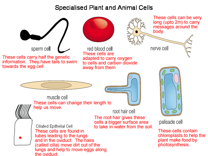
    Complex animals cannot rely on diffusion alone for gas exchange due to their larger body size, lower SA:V ratios, and higher metabolic demands. They require specialised respiratory surfaces and circulatory systems to deliver oxygen efficiently and remove carbon dioxide. Adaptations include gills in fish, tracheal systems in insects, and lungs in mammals. Each system balances efficiency, water conservation, and environmental constraints.

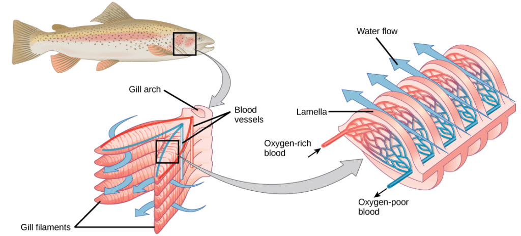
    • Gills are highly folded, thin, and richly supplied with capillaries, providing a large surface area for gas exchange.
    • The countercurrent exchange system maintains a steep concentration gradient: water flows across gill lamellae in the opposite direction to blood flow, maximising oxygen uptake.
    • Gills are adapted for aquatic environments where oxygen is less abundant than in air.
    • Ventilation is maintained by buccal pumping, drawing water continuously over the gill surfaces.

    🧠 Examiner Tip: Always mention countercurrent flow when describing fish gills. Answers that only state β€œwater passes over gills” are incomplete.

    • Insects use a tracheal system where air enters through spiracles and diffuses along tracheae and tracheoles directly to tissues.
    • Tracheoles reach individual cells, reducing diffusion distance.
    • Ventilation is aided by rhythmic body movements that pump air in and out of the tracheae.
    • The tracheal system avoids reliance on blood for oxygen transport, making it highly efficient for small terrestrial animals.

    🧬 IA Tips & Guidance: Students could design investigations into insect spiracle activity, such as observing spiracle opening in response to varying COβ‚‚ concentrations, linking behaviour to efficiency of gas exchange.

    • Mammalian lungs are internal, minimising water loss while allowing efficient gas exchange in a terrestrial environment.
    • The alveoli provide a vast surface area, thin walls, and are surrounded by capillaries, ensuring short diffusion distances and efficient exchange.
    • Surfactant reduces surface tension, preventing alveolar collapse.
    • Oxygen diffuses into red blood cells, binding to haemoglobin for transport, while carbon dioxide diffuses out.

    🌐 EE Focus: An EE could investigate how environmental conditions shape respiratory adaptations β€” for example, comparing fish gill efficiency to mammalian lungs, or examining adaptations in high-altitude animals.

    ❀️ CAS Link: Students could create an educational video comparing respiratory systems across species, then share it with peers or schools to illustrate biological diversity.

    🌍 Real-World Connection: Understanding gas exchange adaptations informs aquaculture (optimising oxygen in fish farming), pest control (targeting insect spiracle activity), and human medicine (treating lung diseases like emphysema).

    • Gills, tracheae, and lungs all maximise surface area, minimise diffusion distance, and maintain concentration gradients.
    • However, strategies differ: fish rely on countercurrent flow, insects use direct diffusion without blood, and mammals use circulatory transport.
    • These systems demonstrate convergent principles of efficiency despite structural differences.

    πŸ” TOK Perspective: Gas exchange systems illustrate how organisms evolve different solutions to the same problem β€” supplying oxygen. TOK questions arise about analogy in science: to what extent can different systems be compared as models of the same principle?

    πŸ“ Paper 2: Paper 2 may ask students to describe and compare gas exchange systems in fish, insects, and mammals. Data-based questions may involve interpreting diagrams of countercurrent exchange or alveolar surface area. Full-mark answers require explaining how each system maintains concentration gradients and short diffusion paths.

  • TermDefinition
    Gas exchangeThe process by which oxygen is absorbed and carbon dioxide is released across a respiratory surface.
    DiffusionThe passive movement of molecules from an area of high concentration to low concentration.
    SA:V ratioThe surface area-to-volume ratio, a key factor determining efficiency of gas exchange.
    Simple diffusionGas exchange directly across a cell membrane without the need for specialised organs.
    Unicellular organismA living organism consisting of a single cell (e.g., Amoeba).

    Single-celled organisms and simple multicellular organisms rely primarily on diffusion across their external surfaces for gas exchange. Their high surface area-to-volume ratio, short diffusion distances, and relatively low metabolic demands allow passive diffusion to meet their oxygen needs and remove carbon dioxide effectively. However, as organisms increase in size and complexity, this method becomes insufficient, necessitating adaptations for more efficient gas exchange.

    • Unicellular organisms such as Amoeba and Paramecium have a very high SA:V ratio, ensuring that diffusion provides sufficient oxygen for cellular respiration.
    • The thin plasma membrane reduces diffusion distance, allowing gases to move in and out rapidly.
    • Their relatively low metabolic demands compared to large animals mean diffusion alone can meet oxygen requirements.
    • Carbon dioxide, produced as a waste product of respiration, diffuses out of the cell along its concentration gradient.
    • Living in aqueous environments facilitates diffusion since gases can dissolve and move efficiently across moist membranes.

    🧠 Examiner Tip: Do not confuse gas exchange (the physical diffusion of gases) with respiration (the chemical process of energy release in cells). This is a common exam error.

    • Flatworms and cnidarians maintain high SA:V ratios due to flattened or simple body plans, enabling gases to diffuse directly across the body surface.
    • Their thin epithelial tissues reduce diffusion distance, further facilitating oxygen uptake.
    • However, increased body size reduces efficiency of simple diffusion, creating a limit on organismal complexity.
    • Many simple animals live in aquatic habitats, as moisture is essential for gas diffusion across membranes.

    🧬 IA Tips & Guidance: Students could investigate diffusion using agar blocks of varying sizes soaked with an indicator such as phenolphthalein, measuring how surface area-to-volume ratio affects diffusion rates β€” a model for constraints on unicellular vs multicellular organisms.

    • As body size increases, SA:V ratio decreases, reducing efficiency of gas exchange across the surface.
    • Protective external structures (e.g., cuticles in some organisms) may reduce permeability.
    • Diffusion alone cannot meet oxygen demands of highly active or large organisms, leading to evolution of specialised respiratory organs.

    🌐 EE Focus: An EE could explore how SA:V ratios affect survival in different environments, for example comparing aquatic vs terrestrial simple organisms, or experimentally modelling diffusion rates in artificial systems of different sizes.

    • Small organisms rely on diffusion due to high SA:V ratio and low metabolic needs.
    • Larger, more active organisms evolve respiratory surfaces and circulatory systems to overcome diffusion limitations.

    ❀️ CAS Link: Students could create models or simple classroom demonstrations of diffusion using coloured dyes in water to illustrate why diffusion is fast in small cells but inadequate in larger bodies.

    🌍 Real-World Connection: Understanding diffusion limitations explains why parasites like flatworms remain small and flat, while more active organisms evolve lungs or gills. It also informs biomedical design, such as drug delivery systems where diffusion constraints must be considered.

    πŸ” TOK Perspective: This topic raises the TOK question: how do physical laws like diffusion limit the possibilities of biology? It shows how scientific models of diffusion not only describe but constrain the evolution of life.

    πŸ“ Paper 2: Paper 2 may ask students to compare gas exchange in unicellular vs multicellular organisms, or to analyse diffusion models (e.g., agar block experiments). Full-mark answers must link SA:V ratio and diffusion distance explicitly to efficiency.

  • TermDefinition
    Specialised cellA cell that has developed specific structures to perform a defined role efficiently.
    AdaptationA structural, physiological, or biochemical feature that enables a cell to perform its function more effectively.
    NeuronA nerve cell specialised for transmitting electrical impulses across long distances.
    Erythrocyte (red blood cell)A cell specialised for oxygen transport via haemoglobin, adapted by lacking organelles.
    Root hair cellA plant cell specialised for absorption of water and minerals from soil.
    Guard cellA specialised plant cell that regulates gas exchange and water loss by controlling stomatal opening.

    Specialised cells embody the principle that β€œform follows function.” Each has evolved structural and biochemical adaptations to optimise its role. For instance, neurons transmit impulses rapidly, while root hair cells maximise absorption. Studying specialised cells demonstrates how organisms achieve efficiency through cellular diversity.

    • Neurons are long and thin, often reaching over a metre in humans, which allows them to transmit impulses rapidly across long distances. Their axons are insulated by myelin sheaths that speed conduction. Synaptic terminals enable communication with multiple target cells.
    • Erythrocytes (red blood cells) are biconcave discs, increasing surface area for gas exchange. They lack nuclei and most organelles, maximising haemoglobin content for oxygen transport. Their flexibility allows them to squeeze through narrow capillaries.
    • Muscle cells are packed with mitochondria to provide ATP for contraction. They contain specialised contractile proteins (actin and myosin) arranged in sarcomeres.
    • Sperm cells have flagella for motility, a midpiece full of mitochondria for energy, and an acrosome containing enzymes to penetrate the egg.

    🧠 Examiner Tip: Always link structural features to function. For example, β€œred blood cells lack a nucleus” is not enough; you must add β€œwhich allows more haemoglobin to be packed inside, increasing oxygen transport.”

    • Root hair cells have elongated extensions that increase surface area for absorbing water and minerals. They contain many mitochondria to fuel active transport of minerals into the root.
    • Guard cells have thickened inner walls and thin outer walls, enabling stomatal opening and closing. They contain chloroplasts to provide ATP for active ion pumping.
    • Xylem vessels are elongated, dead cells with lignified walls that provide structural support and an efficient pathway for water transport.
    • Phloem sieve tube elements lack nuclei and most organelles, allowing efficient transport of sugars, while companion cells maintain metabolic functions.

    🧬 IA Tips & Guidance: Students could investigate how changing environmental conditions (e.g., light intensity) affects guard cell function, linking stomatal behaviour to cell adaptations. Microscopy studies of specialised cells in onion root tips or blood smears also make good practical extensions.

    • Specialised cells illustrate how efficiency results from form-function relationships: neurons optimise communication, erythrocytes optimise transport, and root hairs optimise absorption.
    • This efficiency arises at the cost of flexibility; most specialised cells cannot divide and rely on stem cells for replacement.

    🌐 EE Focus: An EE could compare adaptations of specialised cells across kingdoms, for instance, animal vs plant strategies for maximising surface area (neurons vs root hair cells). Another angle is investigating how structural defects in specialised cells (e.g., sickle cell anaemia in erythrocytes) lead to disease.

    • Specialised cells never function alone β€” tissues and organs depend on their coordination. For example, neurons, muscle cells, and epithelial cells must work together for movement.
    • This interdependence highlights the cooperative nature of multicellularity.

    ❀️ CAS Link: Students could create microscope slide exhibitions of specialised cells, then present them to peers or younger students, linking microscopic features to everyday body and plant functions.

    🌍 Real-World Connection: Many diseases result from failure of specialised cells. For instance, destruction of insulin-producing beta cells in the pancreas causes diabetes, while defective red blood cells cause anaemia. In agriculture, understanding guard cell behaviour aids in developing drought-resistant crops.

    • While specialisation allows high efficiency, it reduces regenerative capacity. For example, neurons rarely divide, making nerve damage difficult to repair.
    • Evolution has balanced this limitation by retaining stem cells for tissue maintenance.

    πŸ” TOK Perspective: Specialised cells raise questions about reductionism versus holism. Looking at a single cell type explains its efficiency, but understanding life requires seeing how diverse cell types integrate into a functioning organism. This echoes TOK discussions of whether breaking systems into parts loses the essence of the whole.

    πŸ“ Paper 2: Paper 2 may require drawing and annotating specialised cells such as root hair cells or red blood cells, or comparing adaptations in animal versus plant cells. Data-based questions often involve interpreting micrographs of specialised tissues. For full marks, students must not only describe features but explicitly explain how each adaptation enables function.

  • TermDefinition
    Cell specialisationThe process by which generic cells develop specific structures and functions suited to a particular role.
    Cell differentiationThe molecular and structural changes that convert a stem cell into a specialised cell type.
    Constraints on cell sizeThe physical and functional limitations that restrict how large or small a cell can be, often linked to surface area-to-volume ratio.
    SA:V ratioThe ratio of surface area to volume, which determines how efficiently a cell can exchange materials with its environment.
    Diffusion limitThe point at which a cell becomes too large for diffusion alone to supply its metabolic needs.

    Cells in multicellular organisms rarely remain identical. Instead, they differentiate into specialised types, enabling the organism to function as an integrated whole. Specialisation improves efficiency but reduces flexibility, meaning cells become committed to particular roles. At the same time, cells face physical limits on their size. The relationship between surface area and volume sets constraints: as cells grow larger, their volume increases faster than their surface area, reducing their ability to exchange materials effectively. These constraints influence why cells remain microscopic, why large organisms consist of many cells rather than fewer giant cells, and why specialisation is necessary for survival.

    • Specialisation allows cells to perform tasks more efficiently than if every cell attempted to perform all functions.
    • Examples include neurons transmitting impulses rapidly, muscle cells contracting for movement, and epithelial cells forming protective barriers.
    • Specialisation is achieved through differential gene expression, where only certain genes are activated in a given cell type, producing unique proteins.
    • The process is irreversible for most cells, locking them into a single role once differentiated.
    • Cell specialisation contributes to division of labour within tissues and organs, enhancing survival and adaptation in complex organisms.

    🧠 Examiner Tip: When explaining cell specialisation, don’t just list examples. Always connect the structural adaptations of a cell to its specific role (e.g., red blood cells lack a nucleus to maximise haemoglobin content for oxygen transport).

    • The SA:V ratio decreases as a cell grows larger, meaning less surface area is available per unit volume for exchange.
    • Smaller cells can exchange gases, nutrients, and wastes more efficiently because of their relatively larger SA:V ratio.
    • If a cell grows too large, diffusion distances become too great, and the centre of the cell may not receive materials quickly enough.
    • Cells overcome this limitation by adopting shapes that maximise surface area (e.g., microvilli in epithelial cells) or by compartmentalisation with organelles.
    • Multicellular organisation evolved partly as a response to this constraint, allowing organisms to grow large while individual cells remain small.

    🧬 IA Tips & Guidance: A classic practical is using agar blocks impregnated with an indicator like phenolphthalein to simulate diffusion in cells. Students can measure how surface area-to-volume ratio affects diffusion rates, linking results to real cellular constraints

    • Flattened or elongated shapes (e.g., red blood cells, nerve cells) increase surface area relative to volume.
    • Internal transport systems such as the cytoskeleton and endomembrane system help move substances efficiently within larger cells.
    • Compartmentalisation within organelles allows localised conditions that improve efficiency despite size.
    • In multicellular organisms, specialised transport systems (e.g., circulatory system) evolve to supply large numbers of cells with nutrients and oxygen.

    🌐 EE Focus: An EE could investigate how different SA:V ratios influence metabolic rates across organisms of different sizes, or compare adaptations to overcome diffusion limits in unicellular vs multicellular organisms.

    • The limits on cell size partly explain why cells differentiate β€” they cannot β€œdo everything” while remaining efficient.
    • Division of labour in tissues offsets size constraints, as no single cell must maintain every function.
    • This balance shows how biology integrates physical limits with evolutionary solutions.

    ❀️ CAS Link: A CAS project could involve designing classroom demonstrations using models of cells with different SA:V ratios, showing younger students why cells remain small and why organisms need specialised cells.

    🌍 Real-World Connection: Cancer cells often lose their specialisation and revert to uncontrolled growth, ignoring normal constraints. Understanding SA:V ratio is also critical in biotechnology, such as designing artificial tissues where nutrient diffusion is a limiting factor.

    • While specialisation enhances efficiency, it reduces flexibility: most specialised cells cannot divide or change role.
    • Stem cells remain unspecialised to balance this limitation, ensuring long-term tissue maintenance.
    • Constraints on cell size reinforce the need for cooperative multicellularity and specialisation.

    πŸ” TOK Perspective: Cell size and specialisation highlight how physical laws (like SA:V ratios) constrain biological possibilities. TOK questions emerge about determinism: to what extent does biology innovate freely, and to what extent is it limited by fundamental physical principles?

    πŸ“ Paper 2: Paper 2 may include data-based questions on SA:V experiments with agar blocks, or ask students to explain why large organisms consist of many small cells rather than fewer large ones. Other questions often compare specialisation in plant and animal cells. To gain full marks, students must explicitly link structure (e.g., cell shape) to function, and relate SA:V constraints to efficiency in exchange.

  • TermDefinition
    Stem cellAn undifferentiated cell capable of unlimited division and differentiation into specialised cells.
    DifferentiationThe process by which stem cells develop into specialised cells with distinct functions.
    PotencyThe capacity of a stem cell to differentiate into different cell types.
    TotipotentStem cells that can form all embryonic and extra-embryonic cell types.
    PluripotentStem cells that can form any embryonic cell type but not extra-embryonic cells.
    MultipotentStem cells that can differentiate into a limited range of closely related cell types.
    UnipotentStem cells that can only divide into their own cell lineage.

    Stem cells are unique because they combine self-renewal with the potential to differentiate into other cell types. This makes them central to embryonic development and to adult tissue repair. Potency defines how versatile a stem cell is β€” totipotent cells have the greatest developmental potential, while unipotent cells are restricted to a single lineage. Understanding stem cells is vital for explaining how multicellular organisms grow, repair, and adapt.

    • Stem cells are maintained in specific β€œniches” β€” microenvironments that control their balance between self-renewal and differentiation.
    • Bone marrow provides a niche for hematopoietic stem cells, enabling the continuous production of red blood cells, white blood cells, and platelets.
    • Hair follicle niches ensure ongoing growth and regeneration of hair.
    • Niches regulate stem cells by keeping them inactive until needed, or stimulating them when repair is required.
    • The environment of the niche provides growth factors, cell-cell signals, and molecular cues that control stem cell fate.

    🧠 Examiner Tip: Always remember the two hallmark properties of stem cells β€” self-renewal and differentiation. In exam answers, link potency terms (totipotent, pluripotent, multipotent, unipotent) to specific examples such as β€œbone marrow stem cells β†’ blood cells.”

    • Totipotent cells (zygote and early embryonic cells up to 16-cell stage) can give rise to all embryonic and extra-embryonic tissues, including placenta.
    • Pluripotent cells (embryonic stem cells) can form any embryonic tissue, but not extra-embryonic tissues.
    • Multipotent cells (adult stem cells, e.g., in bone marrow) can differentiate into a limited but related group, such as all blood cell types.
    • Unipotent cells (e.g., cardiomyocytes) can only divide to produce their own type, but still play a crucial role in tissue maintenance and repair.
    • Potency is gradually lost as development progresses, reflecting increasing specialisation and loss of flexibility.

    🧬 IA Tips & Guidance: Students could design investigations using model organisms such as planarians (flatworms) that demonstrate regeneration through stem cells, linking observations of regrowth to the concept of potency.

    • Embryonic stem cells have immense potential for regenerative medicine, but raise ethical issues.
    • Adult stem cells are used in therapies such as bone marrow transplants for leukaemia.
    • Induced pluripotent stem cells (iPSCs) show how somatic cells can be reprogrammed to pluripotency, offering new therapeutic approaches.

    🌐 EE Focus: An EE could explore the comparative effectiveness of embryonic stem cells versus iPSCs in regenerative therapies, or analyse ethical frameworks surrounding stem cell research

    • Stem cells offer the possibility of replacing damaged tissues in diseases such as Parkinson’s, type I diabetes, or spinal cord injuries.
    • Their ability to self-renew makes them suitable for long-term therapies, unlike transplanted differentiated cells that cannot divide indefinitely.

    ❀️ CAS Link: Students could create informational campaigns or workshops explaining stem cell therapies to the public, engaging in service learning while raising awareness of scientific and ethical issues.

    🌍 Real-World Connection: Stem cells are already used in medicine (bone marrow transplants), and clinical trials continue for conditions like macular degeneration and heart disease. Understanding potency has direct relevance for modern medicine.

    • Stem cell use raises ethical debates, particularly embryonic sources.
    • Scientific advances such as iPSCs aim to resolve ethical dilemmas by avoiding embryo destruction.

    πŸ” TOK Perspective: The study of stem cells highlights tensions between scientific potential and ethical boundaries. It raises the TOK question: should knowledge always be pursued if it has potential benefits, even when ethical objections exist?

    πŸ“ Paper 2: Paper 2 may ask students to define and distinguish totipotent, pluripotent, multipotent, and unipotent cells, often requiring examples. Data-based questions may involve interpreting graphs of stem cell division or experimental results from regenerative research. To gain full marks, answers should connect potency levels to both developmental potential and practical applications.

  • TermDefinition
    RibosomeA molecular machine composed of rRNA and proteins that synthesises polypeptides by translating mRNA.
    Rough Endoplasmic Reticulum (RER)A network of membranes studded with ribosomes, where proteins destined for secretion or membranes are synthesised.
    Signal sequenceA short amino acid sequence that directs ribosomes to the RER for protein targeting.
    Golgi apparatusAn organelle of stacked cisternae that modifies, sorts, and packages proteins into vesicles.
    VesicleA small membrane-bound sac that transports proteins and other molecules within or out of the cell.
    ClathrinA protein that coats vesicles during formation, helping them bud from membranes.

    Protein synthesis and vesicle formation represent one of the most coordinated examples of organelle interaction in eukaryotic cells. Proteins are first produced by ribosomes, either free in the cytoplasm or bound to the rough endoplasmic reticulum. Those intended for secretion, lysosomes, or membranes follow a pathway involving the RER, Golgi apparatus, and vesicular transport. Vesicles act as carriers, moving proteins safely and efficiently through the cytoplasm, ensuring that they reach their correct destination. This system demonstrates compartmentalisation, precision, and regulation, all of which are vital for maintaining cell function.

    • Ribosomes consist of a large and a small subunit, composed of rRNA and protein, which together form the site of translation. They read mRNA codons and assemble amino acids into a polypeptide chain through peptide bond formation.
    • Free ribosomes float in the cytoplasm and primarily synthesise proteins for use inside the cell, such as enzymes in glycolysis or proteins destined for mitochondria and chloroplasts.
    • Bound ribosomes attach to the RER when the polypeptide being synthesised begins with a signal sequence. This sequence directs the ribosome to a receptor on the ER membrane.
    • Once bound to the RER, the ribosome continues translation, and the growing polypeptide chain is threaded into the lumen of the ER, where it may fold and undergo initial modifications.
    • This distinction between free and bound ribosomes reflects compartmentalisation of protein targeting: cytoplasmic use vs secretion or membrane insertion.

    🧠 Examiner Tip: When asked about ribosomes, do not just state β€œthey make proteins.” Specify whether they are free or bound, and always mention the fate of their proteins β€” cytoplasmic use or export/lysosomal pathway.

    • The rough ER provides a surface for ribosome attachment and a lumen where proteins destined for export or membranes enter for folding and modification.
    • Proteins in the RER often undergo glycosylation (addition of carbohydrate groups) or folding with the help of chaperone proteins.
    • From the RER, proteins are packaged into transport vesicles which bud off and travel to the Golgi apparatus.
    • The Golgi consists of flattened sacs called cisternae with a β€œcis” face (receiving side from RER) and a β€œtrans” face (exporting side).
    • As proteins move through the Golgi, they undergo further modifications such as glycosylation, sulfation, or phosphorylation. This ensures that proteins are functional and correctly tagged for their destination.
    • Finally, proteins are sorted into vesicles that deliver them to the plasma membrane, lysosomes, or secretory pathways.

    🧬 IA Tips & Guidance: Students could design investigations using fluorescent protein tagging to track protein movement from ER to Golgi to vesicles. Another practical link is studying enzyme secretion (e.g., amylase) and relating secretion rates to protein synthesis pathways.

    • Vesicles are small, membrane-bound sacs that transport proteins and other macromolecules between organelles or out of the cell.
    • Formation begins with a patch of membrane coated with clathrin proteins, which help bend the membrane into a pit.
    • Receptor proteins on the membrane bind specific cargo molecules, ensuring that vesicles carry only intended contents.
    • Cytoskeletal elements such as actin filaments and microtubules assist in pinching off the vesicle and guiding it to its destination.
    • Vesicles then travel along cytoskeletal tracks using motor proteins like kinesin or dynein, ensuring directed movement.
    • Upon reaching their target, vesicles recognise the correct membrane via β€œSNARE” proteins and fuse, releasing their contents.

    🌐 EE Focus: An EE could explore how vesicle trafficking contributes to diseases such as Alzheimer’s (linked to protein misfolding and mis-targeting) or study how clathrin-mediated endocytosis varies under different experimental conditions. Another angle could investigate the efficiency of protein secretion in plant vs animal cells.

    • The protein pathway demonstrates a coordinated system: DNA β†’ mRNA β†’ ribosome translation β†’ ER modification β†’ Golgi sorting β†’ vesicular transport β†’ final destination.
    • This integration shows how organelles are not isolated but form part of a continuous assembly line, where mistakes at one step (e.g., misfolding in ER) affect the entire chain.
    • Vesicles ensure that proteins remain protected within membranes and are not exposed to cytoplasmic enzymes that could degrade them.
    • Together, protein synthesis and vesicle formation represent one of the clearest examples of structure and function working in harmony within a cell.

    ❀️ CAS Link: A CAS project could involve students making an animation or interactive β€œfactory model” showing ribosomes as workers, ER as assembly lines, Golgi as packaging centres, and vesicles as delivery trucks. Presenting this model in a school outreach program would help simplify complex cellular processes for a wider audience.

    🌍 Real-World Connection: Many diseases are linked to defects in protein synthesis or vesicle trafficking. For example, cystic fibrosis results from misfolded CFTR proteins not reaching the plasma membrane, while lysosomal storage diseases occur when vesicles fail to deliver enzymes properly. In biotechnology, harnessing vesicle pathways is essential for producing and exporting recombinant proteins like insulin.

    • Protein synthesis and vesicle trafficking are highly regulated processes, ensuring that proteins are only made when required and are delivered precisely where needed.
    • Signal sequences and molecular tags (like glycosylation patterns) act as cellular β€œaddresses,” directing proteins to the right location.
    • Vesicle formation is energy-dependent, requiring ATP and GTP hydrolysis, which ensures that transport is tightly controlled and unidirectional.
    • This precise regulation highlights the efficiency and adaptability of eukaryotic cells, which can rapidly increase secretion in response to signals (e.g., hormone release).

    πŸ” TOK Perspective: The pathway of protein synthesis and vesicle trafficking is often taught as a linear model, but in reality, it is highly dynamic, with multiple feedback loops and redundancies. This raises a TOK question: to what extent do simplified models help learning, and when do they risk obscuring the true complexity of biological systems?

    πŸ“ Paper 2: Paper 2 questions on this topic often ask students to describe and explain the pathway of protein synthesis and secretion, starting from ribosomes and ending at the plasma membrane. Data-based questions may include interpreting electron micrographs of RER and Golgi or analysing experimental data on secretion rates. High-mark answers must go beyond naming organelles β€” they should explain how compartmentalisation and vesicle trafficking ensure proteins are efficiently folded, modified, and delivered, showing clear links between structure and function.

  • TermDefinition
    CristaeFolds of the inner mitochondrial membrane that increase surface area for electron transport and ATP synthesis.
    MatrixThe fluid-filled compartment inside mitochondria containing enzymes, DNA, and ribosomes for respiration.
    ThylakoidsFlattened membrane sacs in chloroplasts containing pigments and electron carriers for photosynthesis.
    StromaThe fluid matrix of chloroplasts containing enzymes for the Calvin cycle, DNA, and ribosomes.
    GranaStacks of thylakoids that maximize light absorption for photosynthesis.

    Mitochondria and chloroplasts are specialized double-membrane organelles central to energy transformations. Both show unique structural adaptations that maximize their efficiency in ATP production (mitochondria) and glucose synthesis (chloroplasts). Their compartmentalisation allows separation of biochemical pathways, ensuring rapid and controlled energy conversion.

    • Double membrane system: outer membrane is permeable, inner membrane folded into cristae for large surface area.
    • Cristae host the electron transport chain and ATP synthase.
    • Intermembrane space enables proton accumulation, maintaining a steep gradient essential for oxidative phosphorylation.
    • The matrix contains ribosomes, circular DNA, and enzymes for the Krebs cycle.
    • Compartmentalisation ensures efficient sequencing of respiration (glycolysis β†’ Krebs cycle β†’ oxidative phosphorylation).
    • Active cells (e.g., muscle cells) have more mitochondria with longer, densely packed cristae for higher ATP yield.

    🧠 Examiner Tip: Don’t just state β€œcristae increase surface area.” Always link structure to function: more surface area = more electron carriers + ATP synthase = more ATP production.

    • Double membrane envelope with transport proteins regulates molecule movement.
    • Thylakoid membranes house photosystems, pigments, and electron carriers for the light-dependent stage.
    • Grana stacks maximise surface area for light capture and electron flow.
    • Thylakoid space has very small volume, enabling rapid proton gradient formation.
    • Stroma contains Calvin cycle enzymes, ribosomes, and DNA, supporting protein synthesis for photosynthesis.
    • Photosystems funnel light energy efficiently to reaction centres.

    🧬 IA Tips & Guidance: A practical extension could be comparing oxygen consumption in isolated mitochondria versus starch production in isolated chloroplasts. Such experiments connect organelle structure with measurable activity.

    • Mitochondria: cristae and proton gradients maximise ATP yield.
    • Chloroplasts: grana and thylakoids maximise light capture and carbohydrate synthesis.
    • Both are semi-autonomous, with DNA and ribosomes, supporting endosymbiotic theory.

    🌐 EE Focus: An EE could investigate how chloroplast structures differ between sun and shade plants, or compare mitochondrial density in muscle versus fat tissue, linking structure to metabolic demand.

    • Contain circular DNA and ribosomes β†’ can produce some proteins independently.
    • Evidence for endosymbiotic origin from free-living prokaryotes.
    • Enhances efficiency and adaptability of energy conversion.

    ❀️ CAS Link: Students could build 3D organelle models with removable sections (cristae, grana, stroma) and use them to teach younger students about energy conversion.

    🌍 Real-World Connection: Mitochondrial disorders (e.g., myopathies) result from ATP production failures. In agriculture, chloroplast adaptations are engineered to improve photosynthesis and crop yield.

    • Mitochondria and chloroplasts are interconnected in energy flow.
    • ATP from mitochondria powers biosynthesis, while sugars from chloroplasts fuel respiration.
    • Demonstrates how compartmentalisation supports cooperation between organelles.

    πŸ” TOK Perspective: Focusing on organelles separately is reductionist, but energy efficiency emerges holistically from their integration in the cell. This raises TOK questions on reductionism vs holism in science.

    πŸ“ Paper 2: Paper 2 questions often require linking structure to function, such as explaining how cristae increase ATP production or how grana optimise photosynthesis. Data-based questions may involve interpreting electron micrographs, comparing respiration or photosynthesis rates in isolated organelles, or analysing experimental evidence from cell fractionation. To achieve full marks, answers must not stop at describing structural features but must connect them explicitly to their functional consequences, demonstrating how compartmentalisation enhances efficiency.

  • TermDefinition
    OrganelleA membrane-bound compartment within eukaryotic cells, specialized for particular biochemical processes.
    CompartmentalisationThe separation of cellular activities into distinct organelles or regions within a cell, increasing efficiency and protection.
    LysosomeOrganelle containing hydrolytic enzymes for intracellular digestion and breakdown of waste.
    NucleusOrganelle containing the cell’s genetic material (DNA), controlling gene expression and cell function.
    EndocytosisThe process by which cells engulf external substances by forming vesicles from the plasma membrane.
    Cell fractionationA laboratory technique for separating and studying different organelles by centrifugation.

    Eukaryotic cells differ from prokaryotic cells by their compartmentalised internal structure. Organelles such as the nucleus, mitochondria, endoplasmic reticulum, Golgi apparatus, and lysosomes are enclosed by membranes, which create specialized environments. This organization enables cells to perform complex and often conflicting biochemical reactions simultaneously. Compartmentalisation provides both efficiency and protection, allowing enzymes and substrates to be localized, harmful by-products to be contained, and optimal conditions (pH, concentration) to be maintained for specific processes.

    • Separation of incompatible biochemical reactions (e.g., digestive enzymes in lysosomes are kept away from cytoplasm).
    • Localisation of substrates and enzymes increases reaction efficiency by maintaining high concentrations in small volumes.
    • Maintenance of distinct internal environments, such as low pH in lysosomes or proton gradients in mitochondria.
    • Flexibility: the number and size of organelles can change depending on cellular activity (e.g., muscle cells contain more mitochondria).
    • Compartmentalisation improves regulation, as processes like transcription in the nucleus are separated from translation in the cytoplasm.

    🧠 Examiner Tip: When describing compartmentalisation, always provide examples (e.g., lysosomes preventing self-digestion, or mitochondria isolating respiration steps). Simply stating β€œit increases efficiency” is not enough for full marks.

    • The nucleus is enclosed by a double membrane (nuclear envelope) with pores for exchange of molecules.
    • Separates transcription from translation, reducing the likelihood of errors in mRNA before it meets ribosomes.
    • mRNA can be modified and processed (e.g., splicing, addition of 5’ cap and poly-A tail) before translation.
    • This distinction from prokaryotes highlights the evolutionary advantage of compartmentalisation in maintaining genetic fidelity.

    🧬 IA Tips & Guidance: A practical extension is cell fractionation using centrifugation to isolate organelles. Students can design investigations comparing enzyme activity in lysosomal fractions versus cytoplasm, linking structure to compartmentalisation.

    • Membrane-bound organelles within the cytoplasm (e.g., mitochondria, peroxisomes) keep pathways distinct.
    • Harmful substances can be sequestered safely (e.g., oxidative enzymes in peroxisomes, toxins in vacuoles).
    • In plants, anaerobic niches in the cytoplasm allow sensitive enzymes like nitrogenase to function away from oxygen.
    • Endocytosis and phagocytosis create vacuoles around harmful material, ensuring digestion occurs in controlled compartments.

    🌐 EE Focus: An EE could examine how organelle separation contributes to metabolic efficiency, for example by comparing aerobic respiration in isolated mitochondria vs whole cells. Another angle could be the role of compartmentalisation in evolutionary development of eukaryotes.

    • Early progress in organelle study depended on advances in technology such as electron microscopy and ultracentrifugation.
    • Cell fractionation allows isolation of organelles for biochemical study (e.g., separating mitochondria to investigate respiration).
    • Staining and fluorescent tagging help localise proteins within organelles, linking structure to function.

    ❀️ CAS Link: Students could create models or animations of organelles and their compartmentalised functions, then present these in a community school to explain how cells are organized like β€œfactories with departments.”

    🌍 Real-World Connection: Compartmentalisation explains many disease mechanisms. For example, lysosomal storage disorders (like Tay-Sachs disease) result from enzyme deficiencies within lysosomes. In medicine, targeting drugs to specific organelles (e.g., mitochondria in cancer cells) improves therapy. In biotechnology, compartmentalisation principles inspire nanoreactors and artificial organelles.


    πŸ“Œ Specialisation of Organelles

    • Different organelles evolve adaptations for efficiency: mitochondria for ATP production, chloroplasts for photosynthesis, ER for protein and lipid synthesis.
    • Organelles work together in coordinated pathways (e.g., protein synthesis: ribosomes β†’ ER β†’ Golgi β†’ vesicles β†’ secretion).
    • This cooperation shows that compartmentalisation is not isolation, but controlled interaction.

    πŸ” TOK Perspective: Compartmentalisation highlights how models simplify complex systems. Scientists study organelles separately to understand function β€” but does isolating parts risk missing emergent properties of the whole cell? This raises TOK questions about reductionism versus holism in biology.

    πŸ“ Paper 2: Expect questions comparing compartmentalisation in prokaryotes vs eukaryotes, describing advantages of organelles, and analyzing diagrams. Data-based questions may involve interpreting centrifugation results or enzyme activity in isolated fractions.

  • TermDefinition
    Cell adhesionThe process by which cells attach to each other or to the extracellular matrix, often via specialized proteins.
    Tight junctionA type of cell junction that seals neighboring cells together, preventing leakage of molecules between them.
    DesmosomeStrong, anchoring junctions that link the cytoskeleton of one cell to another, providing mechanical strength.
    Gap junctionChannels that directly connect the cytoplasm of adjacent cells, allowing exchange of ions and small molecules.
    Extracellular matrix (ECM)A network of proteins and polysaccharides outside cells that provides structural and biochemical support.
    IntegrinsTransmembrane proteins that connect the cytoskeleton to the ECM and mediate signaling.

    Cells do not exist in isolation β€” they interact constantly with one another and with their surrounding extracellular environment. These interactions are mediated by membrane proteins, adhesion molecules, and structural networks like the extracellular matrix. Through adhesion and communication, cells form tissues, coordinate activities, and respond to environmental changes. Membrane interactions are essential for maintaining tissue integrity, enabling signaling, and ensuring the cooperative behavior of multicellular organisms.

    • Cells adhere to each other using specialized proteins such as cadherins, integrins, and selectins.
    • Tight junctions form continuous seals between epithelial cells, preventing leakage of materials between compartments (e.g., in the intestines or kidneys).
    • Desmosomes act like rivets, anchoring intermediate filaments between cells and providing resistance to mechanical stress (e.g., in skin and heart muscle).
    • Gap junctions provide direct cytoplasmic connections between neighboring cells, allowing ions and small molecules to pass for rapid communication.
    • These junctions enable tissues to function as integrated units, with both structural and communicative properties.

    🧠 Examiner Tip: In IB exams, when asked about junctions, link structure to function β€” e.g., tight junctions β€œprevent leakage” while gap junctions β€œallow communication.” Avoid simply listing types without explaining their roles.

    • The ECM surrounds animal cells and consists mainly of proteins (collagen, elastin, fibronectin) and polysaccharides (glycosaminoglycans, proteoglycans).
    • Provides structural support, anchoring cells in place, while also serving as a medium for signaling.
    • Integrins connect the ECM to the cytoskeleton, transmitting mechanical and chemical signals that influence cell behavior such as migration, growth, and differentiation.
    • ECM composition varies between tissues, tailoring mechanical and biochemical properties to function (e.g., rigidity in bone vs elasticity in cartilage).

    🧬 IA Tips & Guidance: Investigations using simple histology slides can show tissue differences in ECM structure (cartilage vs muscle). Advanced projects could test enzyme digestion of ECM proteins like collagen, linking structure to functional significance.

    • Membrane proteins act as receptors for hormones, neurotransmitters, and cytokines, enabling cells to detect and respond to external signals.
    • Direct communication occurs through gap junctions in animals and plasmodesmata in plants.
    • Signal transduction pathways often begin at the membrane with receptor-ligand binding, triggering cascades that alter gene expression or metabolism.
    • Effective communication ensures processes like growth, immune response, and tissue repair are coordinated across many cells.

    🌐 EE Focus: An EE could examine how specific adhesion proteins (e.g., integrins or cadherins) influence cancer progression, or how defects in gap junctions affect heart conduction and neurological diseases.

    • Adhesion and membrane interactions allow cells to organize into tissues with distinct functions.
    • In epithelia, tight junctions maintain polarity by separating apical and basal surfaces.
    • In muscle, desmosomes and gap junctions coordinate contraction by linking cytoskeletons and enabling ion flow.
    • The ECM guides cell migration during development and wound healing.
    • Disruption of adhesion leads to diseases such as blistering disorders (loss of desmosomes) or cancer metastasis (loss of anchoring proteins).

    ❀️ CAS Link: Students could design a health-awareness project explaining how cancers spread through loss of adhesion, helping communities understand why early detection and treatment are vital.

    🌍 Real-World Connection: Defects in membrane interactions are implicated in major diseases. For example, loss of cadherins contributes to cancer metastasis, while mutations in gap junction proteins cause heart arrhythmias and deafness. In regenerative medicine, scaffolds mimicking ECM are used to grow replacement tissues. Adhesion molecules are also targeted in therapies for autoimmune diseases and infections.

    • Understanding adhesion helps explain immune responses, where leukocytes bind to blood vessel walls before migrating into tissues.
    • Gap junction studies clarify how heart muscle cells coordinate contraction for pumping blood.
    • ECM research underpins biomedical engineering, leading to artificial cartilage, skin grafts, and tissue scaffolds.
    • Membrane adhesion principles are used in biotechnology to grow cells in culture for research or drug production.

    πŸ” TOK Perspective: Membrane interactions show how biology operates at multiple scales β€” from molecular adhesion to tissue-level behavior. TOK reflection: When studying complex systems like tissues, how do scientists decide the appropriate β€œlevel” of knowledge β€” molecular, cellular, or systemic?

    πŸ“ Paper 2: Be ready to describe tight junctions, desmosomes, and gap junctions; explain roles of ECM and integrins; analyze how adhesion contributes to tissue organization; and apply this knowledge to real-world contexts such as cancer or heart function.

  • TermDefinition
    DiffusionPassive movement of molecules from a region of high concentration to low concentration due to random motion.
    Facilitated diffusionPassive movement of molecules across a membrane through specific channel or carrier proteins.
    OsmosisPassive diffusion of water molecules across a selectively permeable membrane, from low solute concentration to high solute concentration.
    Active transportMovement of molecules against their concentration gradient using energy, typically ATP.
    EndocytosisActive process where the membrane engulfs material to bring it into the cell, forming vesicles.
    ExocytosisActive process where vesicles fuse with the plasma membrane to release contents outside the cell.

    Transport across membranes is vital for maintaining homeostasis, enabling nutrient uptake, waste removal, signal transduction, and energy conversion. Membranes are selectively permeable, meaning only certain molecules can pass freely, while others require assistance. Cells exploit both passive and active processes, as well as bulk transport via vesicles, to control their internal environment. Understanding membrane transport explains phenomena ranging from nerve impulse transmission to kidney filtration.

    • Diffusion is the movement of molecules such as oxygen and carbon dioxide across membranes without energy input. The rate of diffusion depends on concentration gradient, temperature, and surface area.
    • Facilitated diffusion involves large or charged molecules (e.g., glucose, ions) moving through specific protein channels or carriers. It is still passive because it does not require energy.
    • Osmosis is the passive movement of water through aquaporins or directly across the bilayer. Water moves toward regions of higher solute concentration to balance osmotic gradients.
    • Osmosis underlies turgor pressure in plants and fluid balance in animals.

    🧠 Examiner Tip: Always specify the direction of movement relative to solute concentration in osmosis questions, and note that osmosis requires a selectively permeable membrane β€” not just any barrier.

    • Active transport moves molecules against their concentration gradient, requiring energy, usually from ATP hydrolysis.
    • Performed by carrier proteins and pumps (e.g., sodium-potassium pump, proton pumps).
    • The sodium-potassium pump is essential in nerve conduction: it pumps 3 Na⁺ out and 2 K⁺ in, maintaining electrochemical gradients across the membrane.
    • Active transport allows cells to accumulate nutrients, expel waste, and maintain ion gradients essential for processes like respiration and photosynthesis.

    🧬 IA Tips & Guidance: Classic IA experiments include measuring osmosis in potato strips placed in solutions of varying sucrose concentration, or modeling active transport using yeast and glucose uptake with inhibitors to show the need for ATP.

    • Endocytosis: the membrane surrounds and engulfs material, pinching off into vesicles inside the cell. Types include:
      • Phagocytosis (β€œcell eating”) β†’ ingestion of large particles or cells.
      • Pinocytosis (β€œcell drinking”) β†’ uptake of liquids and solutes.
      • Receptor-mediated endocytosis β†’ highly specific, e.g., uptake of LDL cholesterol.
    • Exocytosis: vesicles fuse with the plasma membrane to secrete substances such as hormones, neurotransmitters, or digestive enzymes.
    • Vesicular transport is essential for communication, secretion, and turnover of membrane components.

    🌐 EE Focus: An EE could investigate how temperature, solute concentration, or inhibitors affect transport processes like diffusion or osmosis, or explore vesicular trafficking in relation to diseases like cystic fibrosis.

    • Nerve cells: rely on sodium-potassium pumps, ion channels, and exocytosis of neurotransmitters for impulse transmission.
    • Kidney cells: transport proteins reabsorb glucose, ions, and water from filtrate, demonstrating selective and active transport in physiology.
    • Intestinal cells: glucose absorption occurs via sodium-glucose co-transport, combining facilitated diffusion and active transport.
    • These examples show how universal mechanisms of transport are adapted for specialized functions.

    ❀️ CAS Link: Students could design an experiment-based workshop for younger peers using osmosis in plant tissues (e.g., potato cores in salt solutions) to demonstrate membrane transport, linking school science to nutrition and hydration awareness.

    🌍 Real-World Connection: Many diseases and treatments are based on membrane transport. Cystic fibrosis arises from a defective chloride channel. Diuretics manipulate kidney ion transport to reduce blood pressure. Oral rehydration solutions treat dehydration by exploiting sodium-glucose co-transport. Drug delivery systems often target specific transport mechanisms or vesicular pathways.

    • Explains nutrient uptake in the gut and waste removal by kidneys.
    • Clarifies how plants maintain water balance through osmosis and transpiration.
    • Provides the basis for understanding nerve impulses, muscle contraction, and hormonal secretion.
    • In biotechnology, liposomes and vesicles are engineered for targeted drug delivery.

    πŸ” TOK Perspective: Membrane transport relies on models such as β€œpump” and β€œchannel,” which simplify dynamic molecular processes. TOK reflection: How do metaphors and models in biology aid understanding while also limiting how we perceive reality?

    πŸ“ Paper 2: Be ready to distinguish passive vs active transport, explain sodium-potassium pump operation, describe endocytosis and exocytosis with diagrams, and interpret experimental data on osmosis and diffusion rates.