B2.1.1 – MEMBRANE STRUCTURE
📌Definition Table
| Term | Definition |
|---|---|
| Amphipathic | Molecule with both hydrophilic and hydrophobic regions. |
| Phospholipid | Lipid with two fatty acids and a phosphate group attached to glycerol. |
| Integral Protein | Membrane protein embedded within the lipid bilayer, often spanning the membrane. |
| Peripheral Protein | Membrane protein loosely bound to the surface of the membrane. |
| Glycolipid | Lipid with carbohydrate chain attached, involved in cell recognition. |
| Fluid Mosaic Model | Model describing the membrane as a dynamic structure with proteins embedded in a fluid lipid bilayer. |
📌Introduction
The plasma membrane is a selectively permeable barrier that controls the entry and exit of substances, enabling compartmentalisation and communication. Its amphipathic phospholipid bilayer forms the basic structure, with embedded proteins, cholesterol, and carbohydrates providing additional functions.
❤️ CAS Link: Lead a microscopy demonstration for junior students, showing onion epidermis or cheek cells stained to highlight membranes.
📌 Lipid Bilayer and Amphipathic Nature
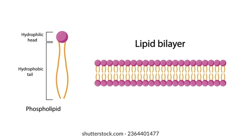
- Composed of phospholipids arranged with hydrophilic heads outwards and hydrophobic tails inwards.
- Amphipathic nature creates a barrier to polar molecules and ions.
- Bilayer provides flexibility and allows for self-healing.
- Fatty acid composition (saturated vs unsaturated) affects fluidity.
- Cholesterol interspersed in the bilayer moderates fluidity — reduces movement at high temperatures, prevents solidification at low temperatures.
- Membrane asymmetry — different lipid composition in inner vs outer leaflet.
🧠 Examiner Tip: Always connect phospholipid amphipathic nature to selective permeability in long-answer questions.
📌 Membrane Proteins
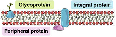
- Integral proteins: Span the bilayer, often involved in transport, receptors, and enzyme activity.
- Peripheral proteins: Attached to membrane surface; often act in cell signalling or as structural anchors.
- Transport proteins: channel proteins (passive transport) and carrier proteins (active/facilitated transport).
- Receptor proteins: bind to specific signalling molecules, triggering responses.
- Enzymatic proteins: catalyse reactions at the membrane surface.
- Adhesion proteins: help cells stick together in tissues.
🌍 Real-World Connection: Mutations in membrane transport proteins can cause diseases such as cystic fibrosis.
📌 Carbohydrates in Membranes
- Glycolipids: Lipid + carbohydrate chain; aid in cell recognition and stability.
- Glycoproteins: Protein + carbohydrate chain; function in signalling, immune recognition, and adhesion.
- Carbohydrate chains extend outward into the extracellular space forming the glycocalyx.
- Important in tissue formation and immune defence.
- Pathogens may exploit glycocalyx for attachment to host cells.
- Serve as antigens in blood groups (A, B, AB, O).
🌐 EE Focus: Investigate the role of glycoproteins in pathogen binding using microscopy or biochemical assays.
📌 Fluid Mosaic Model
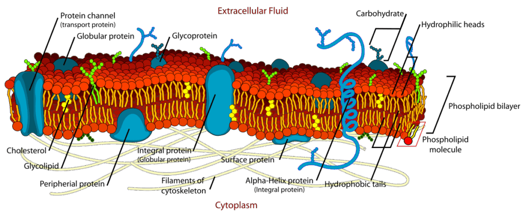
- Proposed by Singer & Nicolson (1972).
- Membrane is a fluid lipid bilayer with proteins embedded, giving a mosaic appearance.
- Components are dynamic — lateral movement of lipids and proteins.
- Explains membrane flexibility, repair, and transport function.
- Supported by freeze-fracture electron microscopy evidence.
- Protein mobility can be restricted by cytoskeletal attachments.
🔍 TOK Perspective: The shift from the Davson-Danielli model to the Fluid Mosaic Model highlights how new evidence can overturn long-accepted scientific models.