A2.2.3 – COMPARISON AND MICROSCOPY
📌Definition Table
| Term | Definition |
|---|---|
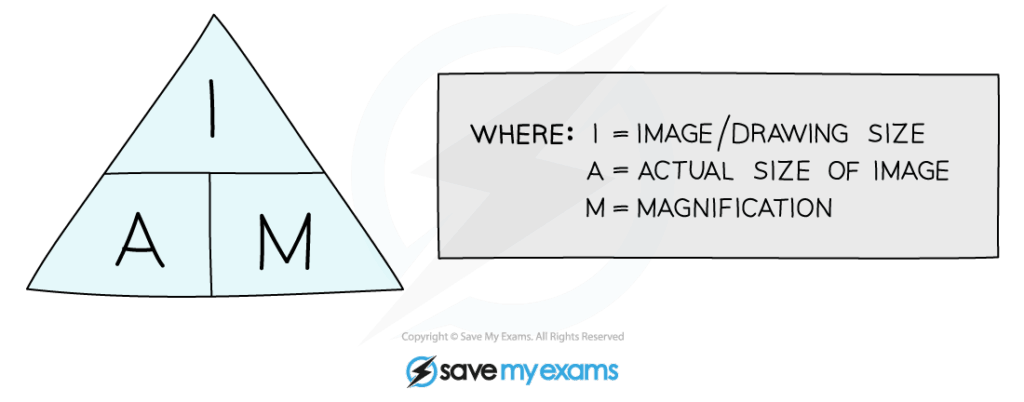
| Magnification | The number of times an image is enlarged compared to its actual size. |
| Resolution | The ability of a microscope to distinguish two points as separate. |
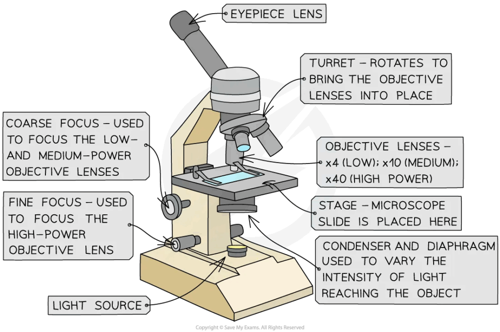
| Light Microscope | Microscope using visible light and lenses to view specimens. |
| Electron Microscope | Microscope using beams of electrons to achieve higher resolution. |
| Scanning Electron Microscope (SEM) | Electron microscope producing 3D surface images. |
| Transmission Electron Microscope (TEM) | Electron microscope producing 2D images of thin specimens. |
📌Introduction
Comparing cell types and understanding microscopy techniques are essential for studying cell ultrastructure. Key differences between prokaryotic and eukaryotic cells, and between plant and animal cells, help explain how structural features relate to function. Microscopy allows these features to be visualised in detail, with advances in technology significantly improving our ability to study cells.
❤️ CAS Link: Organise a microscopy workshop for younger students, where they can observe plant and animal cells and learn how to calculate magnification from scale bars.
📌 Prokaryotic vs Eukaryotic Cells
- Prokaryotic cells are smaller (0.1–5 μm) and lack membrane-bound organelles; eukaryotic cells are larger (10–100 μm) and have organelles.
- DNA in prokaryotes is circular and located in the nucleoid; in eukaryotes it is linear and enclosed in a nucleus.
- Prokaryotes have 70S ribosomes, eukaryotes have 80S ribosomes (and 70S in mitochondria/chloroplasts).
- Prokaryotes reproduce by binary fission; eukaryotes by mitosis and meiosis.
- Prokaryotic cell walls contain peptidoglycan; eukaryotic plant cell walls contain cellulose.
- Prokaryotes generally lack cytoskeletal structures, while eukaryotes have a complex cytoskeleton.
🧠 Examiner Tip: In “compare and contrast” questions, always balance similarities and differences — unbalanced answers can lose marks.
📌 Plant vs Animal Cells
- Plant cells have a rigid cell wall, large central vacuole, and chloroplasts; animal cells do not.
- Animal cells have centrioles for cell division; plant cells generally do not.
- Plant cells store starch; animal cells store glycogen.
- Lysosomes are more common in animal cells.
- Plasmodesmata allow communication between plant cells; gap junctions serve this role in animal cells.
- Both plant and animal cells have a plasma membrane, mitochondria, ER, Golgi apparatus, and ribosomes.
🌍 Real-World Connection: Understanding plant–animal cell differences is essential in biotechnology, such as tissue culture and crop genetic engineering.
📌 Light Microscopy vs Electron Microscopy
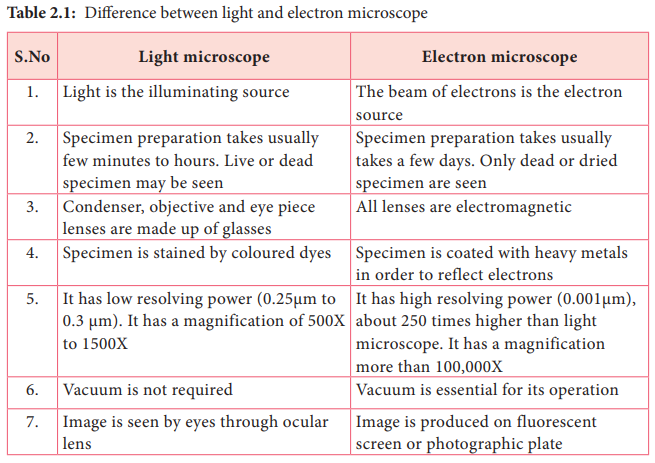
- Light microscopes use visible light and glass lenses; resolution ~200 nm.
- Electron microscopes use electron beams; resolution ~0.1 nm.
- TEM produces 2D images of thin specimens, showing internal structures.
- SEM produces 3D images of surfaces.
- Electron microscopy requires specimens to be in a vacuum and coated in metal.
- Light microscopy can be used for living cells; electron microscopy cannot.
🔍 TOK Perspective: The invention of the electron microscope transformed biology — but does increasing resolution always increase understanding?
📌 Magnification and Resolution Calculations
- Magnification formula: Image size ÷ Actual size.
- Units must be converted before calculation (e.g., μm to mm).
- Resolution depends on the wavelength of light/electrons — shorter wavelengths give higher resolution.
- Scale bars on micrographs help determine magnification and actual size.
- Proper calculation requires significant figures consistent with data given.
- IB often tests both calculation and interpretation of micrographs.
⚗️ IA Tips & Guidance: A microscopy IA could compare light vs electron microscope resolution using prepared images or measure the size of organelles in micrographs.