B3.3.2 – MECHANISM OF MUSCLE CONTRACTION
📌Definition Table
| Term | Definition |
|---|---|
| Sarcomere | The functional unit of a myofibril, defined as the region between two Z-lines. |
| Actin | Thin filament protein that forms the backbone of the sarcomere; interacts with myosin for contraction. |
| Myosin | Thick filament protein with heads that bind actin, hydrolyze ATP, and generate movement. |
| Tropomyosin | Protein that covers actin binding sites in resting muscle. |
| Troponin | Regulatory protein that binds calcium ions, causing tropomyosin to shift and expose actin sites. |
| Sliding filament theory | Model describing contraction as actin filaments sliding over myosin filaments, shortening the sarcomere. |
📌Introduction
Muscle contraction underlies all active movement in animals, from heartbeat to locomotion. The basic mechanism is explained by the sliding filament theory, where actin and myosin filaments interact within the sarcomere. The process is powered by ATP and regulated by calcium ions released from the sarcoplasmic reticulum. Contraction transforms chemical energy into mechanical force, a unifying principle across muscle types (skeletal, cardiac, smooth).
📌 Sarcomere Structure
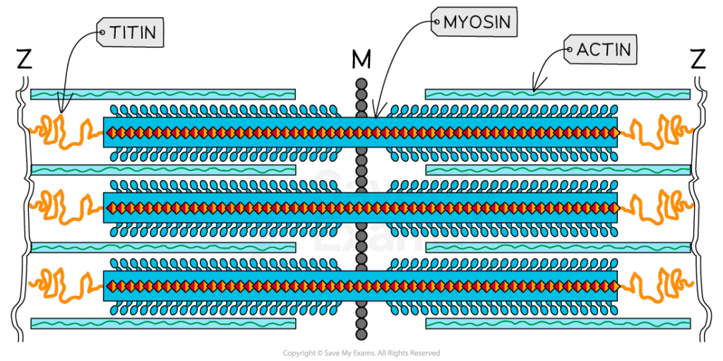
- Each sarcomere is bounded by Z-lines, anchoring actin filaments.
- A band: dark band containing overlapping actin and myosin filaments.
- I band: light band containing actin only.
- H zone: central region with myosin only.
- During contraction:
- Sarcomere shortens, Z-lines move closer.
- I band and H zone shrink, but A band remains constant.
- This structural change is observable under electron microscopy.
🧠 Examiner Tip: Examiners often test sarcomere changes. Remember: A band stays the same, while I band and H zone shorten.
📌 Cross-Bridge Cycle
- Resting state: tropomyosin blocks actin binding sites.
- Excitation: nerve impulse triggers acetylcholine release, depolarizing the sarcolemma.
- Calcium release: depolarization spreads via T-tubules, stimulating sarcoplasmic reticulum to release Ca²⁺.
- Binding: Ca²⁺ binds troponin, shifting tropomyosin and exposing actin sites.
- Cross-bridge formation: myosin heads bind actin, forming cross-bridges.
- Power stroke: myosin head pivots, pulling actin toward the M line, releasing ADP + Pi.
- Detachment: ATP binds myosin, releasing it from actin.
- Resetting: ATP hydrolysis re-cocks the myosin head.
- The cycle repeats as long as Ca²⁺ and ATP are present.
🧬 IA Tips & Guidance: Students can model the sliding filament theory with physical props (sticks and hooks) or use bioinformatics tools to analyze muscle protein structures. Physiological labs can involve measuring muscle fatigue under repeated stimulation.
📌 Role of ATP and Calcium
- ATP functions:
- Powers myosin head movement (power stroke).
- Breaks cross-bridges by binding myosin.
- Powers Ca²⁺ reuptake into sarcoplasmic reticulum.
- Calcium ions:
- Act as the trigger by binding troponin.
- Maintain contraction as long as they remain in cytosol.
- Removal of Ca²⁺ leads to relaxation.
🌐 EE Focus: An EE could explore how ATP availability or calcium ion concentration affects contraction efficiency. Topics could include muscle fatigue in high-intensity exercise or comparing calcium regulation in skeletal vs cardiac muscle.
📌 Neuromuscular Junction
- Motor neurons release acetylcholine (ACh) into synaptic cleft.
- ACh binds receptors on sarcolemma, opening sodium channels and depolarizing the muscle fiber.
- Action potential spreads along sarcolemma and into T-tubules.
- Ensures rapid and coordinated contraction of the muscle fiber.
- Acetylcholinesterase breaks down ACh, resetting the system.
❤️ CAS Link: Students could organize workshops showing how reaction time and reflexes involve neuromuscular coordination, linking sports science and biology to community fitness or safety programs.
🌍 Real-World Connection:
Understanding contraction underpins treatment of neuromuscular disorders (e.g., myasthenia gravis, muscular dystrophy). Drugs and toxins (e.g., curare, botulinum toxin) target neuromuscular junctions. Sports physiology applies knowledge of muscle metabolism and fatigue to improve training. Robotics and prosthetics mimic sliding filament mechanics in artificial actuators.
📌 Integration with Muscle Types
- Skeletal muscle: voluntary, rapid, fatigue-prone; multinucleate and striated.
- Cardiac muscle: involuntary, striated, highly resistant to fatigue due to many mitochondria and intercalated discs.
- Smooth muscle: involuntary, non-striated; slower contractions for long-term control (e.g., peristalsis).
- Despite differences, all rely on actin–myosin interactions and ATP hydrolysis.
🔍 TOK Perspective: Muscle contraction is studied using reductionist models (sarcomere isolated under microscopes). TOK reflection: How much of biological understanding is lost when studying systems in isolation rather than in the whole organism?