A2.2.1 – PROKARYOTIC CELL STRUCTURE AND FUNCTION
📌Definition Table
| Term | Definition |
|---|---|
| Prokaryote | Single-celled organism lacking a membrane-bound nucleus and organelles. Includes Bacteria and Archaea. |
| Plasmid | Small, circular DNA molecule separate from chromosomal DNA, often carrying extra genes like antibiotic resistance. |
| Nucleoid | Region in a prokaryotic cell where the circular DNA is located, not enclosed by a membrane. |
| Pilus (pili) | Hair-like appendages on prokaryotes used for attachment or DNA transfer during conjugation. |
| Binary Fission | Asexual reproduction process in prokaryotes where the cell divides into two identical cells. |
📌Introduction
Prokaryotic cells are the most ancient and structurally simple forms of life, appearing on Earth over 3.5 billion years ago. They lack a true nucleus and membrane-bound organelles, yet they perform all essential life functions such as metabolism, growth, and reproduction. Their compact and efficient structure allows them to thrive in diverse environments, from deep-sea vents to the human gut. Understanding prokaryotic cell structure is key to microbiology, biotechnology, and medicine, as many prokaryotes are either beneficial (e.g., nitrogen-fixing bacteria) or harmful (pathogens).
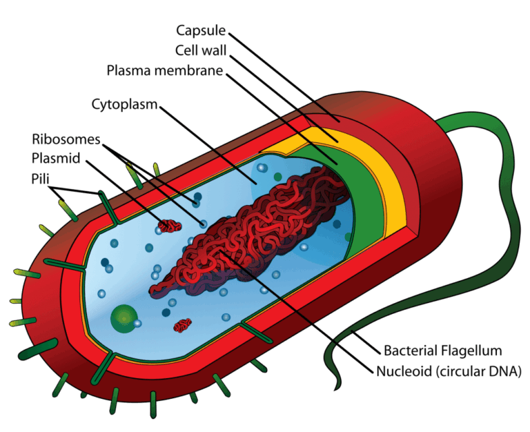
📌 Structure of Prokaryotic Cells
- Cell wall composed of peptidoglycan (in Bacteria) or other polymers (in Archaea), providing shape and protection.
- Plasma membrane controls movement of substances in and out of the cell.
- Nucleoid contains a single, circular DNA molecule, the main genetic material.
- Plasmids carry extra genes, often conferring survival advantages.
- Ribosomes (70S) are the sites of protein synthesis.
- Flagella enable motility, powered by a rotary motor mechanism.
- Pili and fimbriae assist in attachment to surfaces and in conjugation.
🧠 Examiner Tip: Always specify 70S ribosomes when describing prokaryotic cells in IB exams, as it’s a common marking point.
📌 Reproduction & Gene Transfer
- Prokaryotes reproduce asexually via binary fission, producing genetically identical cells.
- DNA is replicated starting at a single origin of replication before cell division.
- Conjugation allows DNA transfer between prokaryotes via pili.
- Transformation occurs when prokaryotes absorb foreign DNA from the environment.
- Transduction uses viruses (bacteriophages) to transfer genetic material between cells.
- These methods contribute to rapid adaptation and evolution in bacterial populations.
🧬 IA Tips & Guidance: For a lab investigation, bacterial growth curves can be measured under different environmental conditions, linking cell structure to survival.
📌 Specialized Structures in Some Prokaryotes
- Capsule: protective layer preventing desiccation and aiding immune evasion.
- Endospores: dormant, resistant structures for surviving extreme conditions.
- Thylakoid membranes in cyanobacteria for photosynthesis.
- Magnetosomes for orientation in magnetic fields.
- Gas vesicles for buoyancy control in aquatic environments.
- Plasma membrane infoldings to increase surface area for metabolic processes.
🌐 EE Focus: An EE could explore structural adaptations of extremophile prokaryotes and how these allow survival in extreme environments.
📌 Functions of Prokaryotic Cells
- Maintain homeostasis through selective permeability of the plasma membrane.
- Perform metabolism, including respiration, fermentation, and photosynthesis (in some species).
- Protect against environmental stress through cell wall and capsule formation.
- Engage in symbiotic relationships (e.g., gut microbiota in humans).
- Adapt rapidly to environmental change via high mutation rates and horizontal gene transfer.
- Act as decomposers, nitrogen fixers, and producers in ecosystems.
❤️ CAS Link: A CAS project could involve creating public awareness materials on antibiotic resistance, linking bacterial gene transfer to public health.
🌍 Real-World Connection:
Knowledge of prokaryotic cell structures underpins antibiotic development, as drugs often target specific bacterial components like the cell wall or ribosomes.
📌 Microscopy in Prokaryotic Studies
- Light microscopes allow observation of general shape and arrangement of cells.
- Electron microscopes reveal internal details, such as ribosomes and nucleoid structure.
- Staining techniques (Gram staining) differentiate bacterial cell wall types.
- Fluorescence microscopy can highlight specific proteins or DNA sequences.
- Time-lapse microscopy can show binary fission in real time.
- Microscopy is essential for taxonomy, pathology, and research into cell function.
🔍 TOK Perspective: Our understanding of prokaryotic cells depends heavily on technological advancements in microscopy — without these tools, much of modern microbiology would not exist.