A2.2.3 – COMPARISON AND MICROSCOPY
📌Definition Table
| Term | Definition |
|---|---|
| Resolution | The ability of a microscope to distinguish two points as separate. |
| Magnification | The process of enlarging an image compared to the actual size of the specimen. |
| Gram Staining | A method to classify bacteria based on cell wall composition. |
| Light Microscope (LM) | Microscope that uses visible light to magnify images, suitable for living specimens. |
| Electron Microscope (EM) | Uses electron beams for much higher resolution images, requires dead specimens. |
📌Introduction
Comparing prokaryotic and eukaryotic cells reveals key differences in complexity, structure, and evolutionary history. Prokaryotes are smaller, lack membrane-bound organelles, and have circular DNA, while eukaryotes are larger, contain multiple organelles, and have linear chromosomes within a nucleus. These differences are closely linked to their functions, reproductive methods, and ecological roles. Microscopy — from simple light microscopes to advanced electron microscopes — has been the key tool in uncovering these distinctions, enabling scientists to study cell ultrastructure in detail and validate theories such as endosymbiosis.
📌 Prokaryotic vs Eukaryotic Differences
- Size: Prokaryotic cells are typically 0.1–5 μm; eukaryotic cells are 10–100 μm.
- DNA: Prokaryotes have a single circular chromosome in a nucleoid; eukaryotes have multiple linear chromosomes in a nucleus.
- Ribosomes: Prokaryotic ribosomes are 70S; eukaryotic cytoplasmic ribosomes are 80S.
- Organelles: Prokaryotes lack membrane-bound organelles; eukaryotes have many specialised ones.
- Reproduction: Prokaryotes divide by binary fission; eukaryotes divide by mitosis or meiosis.
- Cell wall: Present in most prokaryotes (peptidoglycan in bacteria) and some eukaryotes (cellulose in plants, chitin in fungi).
🧠 Examiner Tip: IB questions often award marks for correct, concise comparison tables of prokaryotic vs eukaryotic structures — memorise at least three clear differences.
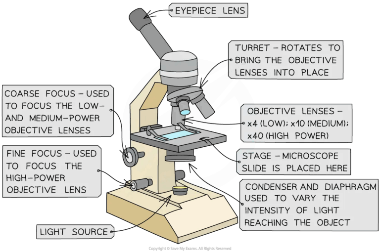
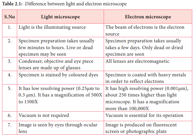
📌 Light Microscopy
- Uses visible light to illuminate the specimen.
- Magnification up to ~1000× with resolution of ~200 nm.
- Suitable for living specimens, stained slides, and dynamic processes.
- Common stains: methylene blue, iodine, crystal violet.
- Inexpensive and accessible for most labs.
- Limited in resolving very small structures like ribosomes.
🧬 IA Tips & Guidance: Light microscopes are excellent for IA work — choose appropriate stains and measure field of view to calculate actual specimen size.
📌 Electron Microscopy
- Transmission Electron Microscope (TEM) passes electrons through the specimen for internal details at ~0.1 nm resolution.
- Scanning Electron Microscope (SEM) scans specimen surface to produce 3D images.
- Requires specimens to be fixed, dehydrated, and coated in metals (gold, platinum).
- Cannot be used on living specimens.
- Essential for viewing organelles like mitochondria, ER, and ribosomes in detail.
- More costly and requires specialised training.
🌐 EE Focus: An EE could investigate how resolution differences between light and electron microscopy influence the discovery of cellular structures.
📌 Fluorescence and Confocal Microscopy
- Uses fluorescent dyes or proteins (e.g., GFP) to highlight specific cell components.
- Confocal microscopy uses laser scanning to produce sharp, 3D reconstructions.
- Allows visualisation of dynamic processes in living cells.
- Common in molecular biology and medical research.
- Can be combined with electron microscopy for correlative studies.
- Offers greater specificity than traditional staining methods.
❤️ CAS Link: A CAS project could involve creating microscopy workshops for younger students, showing how to prepare slides and interpret images.
📌 Importance of Microscopy in Cell Theory
- Discovery of cells in the 17th century relied on light microscopy.
- Electron microscopy in the 20th century revealed organelles and ultrastructure.
- Led to the formulation and refinement of cell theory.
- Validated endosymbiotic theory by showing bacterial-like features in mitochondria/chloroplasts.
- Continues to drive research in cell biology, microbiology, and nanotechnology.
- Demonstrates how technology influences scientific progress.
🔍 TOK Perspective: Microscopy shows how technological limitations shape what we can know — improvements often lead to paradigm shifts in biology.
🌍 Real-World Connection:
Microscopy is critical in medical diagnostics, allowing detection of pathogens, cancerous cells, and tissue abnormalities at an early stage.